Quantcast
Channel: Airyscan – Microscopy
Viewing all 43 articles
Browse latest View live

ZEISS Microscopes Help to Link Heart Disease, Leukemia to Dysfunction in Nucleus

$
0
0

Salk scientists identify the cell nucleus as a driver of gene expression and, sometimes, disease

Salk scientists identify the cell nucleus as a driver of gene expression and, sometimes, disease. Arkaitz Ibarra and Martin Hetzer with ZEISS confocal microscope systems. Credit: Salk Institute

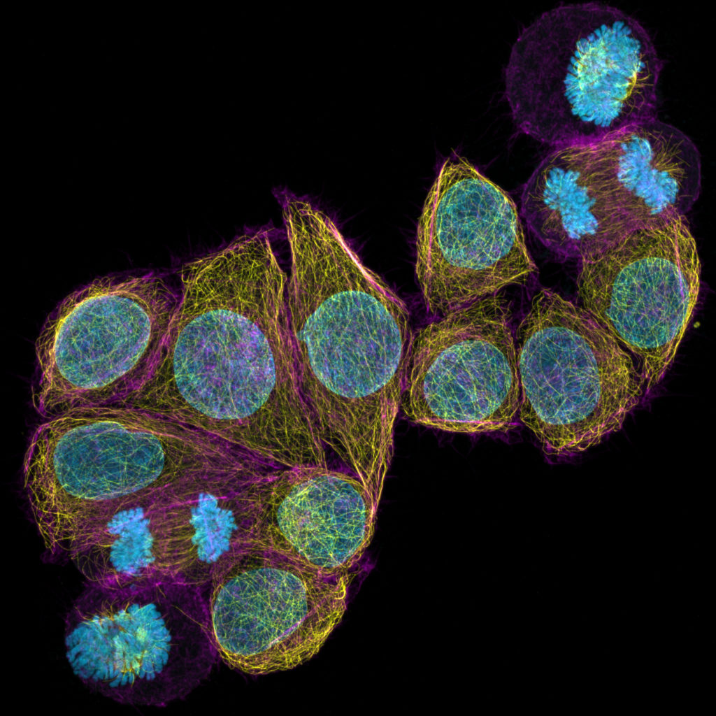
We put things into a container to keep them organized and safe. In cells, the nucleus has a similar role: keeping DNA protected and intact within an enveloping membrane. But a new study by Salk Institute scientists, detailed in the November 2 issue of Genes & Development, reveals that this cellular container acts on its contents to influence gene expression. Microscopy was performed with a ZEISS LSM confocal microscope system. “Our research shows that, far from being a passive enclosure as many biologists have thought, the nuclear membrane is an active regulatory structure,” says Salk Professor Martin Hetzer, who is also holder of the Jesse and Caryl Philips Foundation chair. “Not only does it interact with portions of the genome to drive gene expression, but it can also contribute to disease processes when components are faulty.”

Using a suite of molecular biology technologies, the Salk team discovered that two proteins, which sit in the nuclear envelope, together with the membrane-spanning complexes they form, actively associate with stretches of DNA to trigger expression of key genes. Better understanding these higher-level functions could provide insight into diseases that appear to be related to dysfunctional nuclear membrane components, such as leukemia, heart disease and aging disorders. Historically, the nuclear membrane’s main purpose was thought to be keeping the contents of the nucleus physically separated from the rest of the cell. Complexes of at least thirty different proteins, called nucleoporins, form gateways (pores) in the membrane, controlling what goes in or out. But as the Hetzer lab’s work on nucleoporins shows, these nuclear pore complexes (NPCs), beyond being mere gateways into the nucleus, have surprising regulatory effects on the DNA inside.

Salk scientists discover that nuclear pore components regulate the expression of cell identity genes through functional interactions with super-enhancers. In the image, a super-enhancer driven cell identity gene (red dot) localizes in close proximity to the nuclear envelope (green) in the nucleus of human primary lung fibroblasts (blue). Credit: Salk Institute

“Discovering that key regulatory regions of the genome are actually positioned at nuclear pores was very unexpected,” says Arkaitz Ibarra, a Salk staff scientist and first author of the paper. “And even more importantly, nuclear pore proteins are critical for the function of those genomic sites.” Curious about all the regions of DNA with which nucleoporins potentially interact, the team turned to a human bone cancer cell line. The scientists used a molecular biology technique called DamID to pinpoint where two nucleoporins, Nup153 and Nup93, came into contact with the genome. Then they used several other sequencing techniques to understand which genes were being affected in those regions, and how. The Salk team discovered that Nup153 and Nup93 interacted with stretches of the genome called super-enhancers, which are known to help determine cell identity. Since every cell in our body has the same DNA, what makes a muscle cell different from a liver cell or a nerve cell is which particular genes are turned on, or expressed, within that cell. In the Salk study, the presence of Nup153 and Nup93 was found to regulate expression of super-enhancer driven genes and experiments that silenced either protein resulted in abnormal gene expression from these regions. Further experiments in a lung cancer cell line validated the bone cancer line results: Nucleoporins in the NPC were found to interact with multiple super-enhancer regions to drive gene expression, while experiments that altered the NPC proteins made related gene expression faulty, even though the proteins still performed their primary role as gatekeepers in the cell membrane.

“It was incredible to find that we could perturb the proteins without affecting their gateway role, but still have nearby gene expression go awry,” says Ibarra. The results bolster other work indicating that problems with the nuclear membrane play a role in heart disease, leukemia and progeria, a rare premature aging syndrome. “People have thought the nuclear membrane is just a protective barrier, which is maybe the reason why it evolved in the first place. But there are many more regulatory levels that we don’t understand. And it’s such an important area because so far, every membrane protein that has been studied and found to be mutated or mis-localized, seems to cause a human disease,” says Hetzer.

Other authors on the paper were Swati Tyagi of the Salk Institute, Chris Benner of the University of California, San Diego, and Jonah Cool of Organovo Holdings, Inc.

Genes & Development: Nucleoporin-mediated regulation of cell-identity genes; Arkaitz Ibarra, Chris Benner, Swati Tyagi, Jonah Cool and Martin W. Hetzer

The ZEISS LSM 8 family of confocal microscopes with Airyscan: Revolutionize your confocal imaging! 

Learn more in the Salk Institute’s video about the work of Martin Hetzer and his group

Article originally appeared at the Salk Institute news blog. Text & images courtesy of the Salk Institute, Office of Communications, 2016. The work was funded by the Human Frontier Science Program, National Institutes of Health grant R01GM098749, NIH Transformative Research Award R01NS096786, the Glenn Foundation For Medical Research, the NOMIS Foundation, the Keck Foundation and American Cancer Society Award number P30CA014195.

The post ZEISS Microscopes Help to Link Heart Disease, Leukemia to Dysfunction in Nucleus appeared first on Microscopy News Blog.


Report from Neuroscience 2016 in San Diego

$
0
0

ZEISS shows new Celldiscoverer 7 and Axio Observer, wins #MyNeuroVote people's choice award third time in a row

The ZEISS team at Neuroscience 2016

This year over 30.000 people attended the 46th annual meeting of the Society for Neuroscience (SfN) at the San Diego Convention Center. From November 12-16, ZEISS joined the premier venue for neuroscientists to present our newest instruments and technologies. 3D imaging in light, electron and X-ray microscopy is essential to decipher the human connectome, assist in brain research, or help to fight neurodegenerative diseases such als Alzheimer’s, and ZEISS once again demonstrated the perfect tools and applications to enthusiastic scientists.

Professor Jochen Herms, LMU Munich, presenting correlative microscopy to a full audience at the ZEISS booth

Visitors had the chance to discover the latest ZEISS advancements in confocal and multi-photon microscopy, rapid imaging of large cleared samples and correlative scanning electron microscopy. The ZEISS booth seminars by customers and experts included insights into new voltage-sensitive dyes for ex vivo brain imaging, correlative in vivo 3D multiphoton and electron microscopy, high-throughput connectomics with MultiSEM technology, and expansion microscopy with ZEISS Lightsheet Z.1.

Crowd magnet: arivis immersive VR microscopy experience with gesture control and real 3D datasets at Neuroscience 2016

ZEISS and arivis for the first time presented the world-exclusive premier of the arivis InViewR, a 3D visualization of real microscopy data with Oculus Rift virtual reality technology and Leap Motion gesture controls. Excited visitors took the chance to dive deep into real 3D microscopy data, interact with the 3D datasets by using their own hands, and experience immersive microscopy first-hand.

Automated microscopy, made by ZEISS: Axio Scan.Z1 & Celldiscoverer 7

Available instruments for hands-on demonstrations included our new Axio Observer family, Axio Scan.Z1, the LSM 8 family of confocal microscopes with Airyscan, ZEISS GeminiSEM with ATUMtome array tomography, Lightsheet Z.1 for clearing and expansion microscopy, and our digital classroom experience. A special highlight was the world premiere of ZEISS Celldiscoverer 7, the new automated platform for live cell imaging.

ZEISS LSM 880 Airyscan Fast Mode wins #MyNeuroVoteA very special honour was to receive the #MyNeuroVote people’s choice award for LSM 880 with Airyscan in the third year running. Johannes Amon, of ZEISS Microscopy, told SelectScience that it was a an honor to be selected by researchers in the community. “Winning the award for the third time in a row shows the acceptance and validation of this innovative product. #MyNeuroVote is the scientists’ choice award for Neuroscience, and ZEISS LSM 880 is a confocal microscopy platform that continues to propel our customers’ research forward. With the new Fast Mode we demonstrate that we are still extending the possibilities of the unique Airyscan detector technology. The continuous flow of awards shows us that Airyscanning is recognized as a truly revolutionary confocal technology compared to competing technologies that are merely software-based.” Our whole team at ZEISS Microscopy would like to thank all Neuroscience 2016 participants for visiting our booth and for voting! We had great discussions, enjoyed the instruments demonstrations, learned about your applications, and had a great and successful time together with you.


We are looking forward meeting you again at Neuroscience 2017 in Washington, DC! In case you have more questions, comments, or are interested in the presented instruments and applications, just write us an email at microscopy@zeiss.com or use our web contact form: We’re always there for you!

Next stop for our team and the new instruments is the ZEISS booth at the ASCB meeting in San Francisco from December 4-7. See you there!

A young researcher at Neuroscience 2016, impressed by ZEISS LSM 800 with Airyscan

Our favourite moments from the show are collected on Storify: Enjoy the impressions!

Want so see more photos from the show? Visit our flickr album!

Can’t see the embedded video? Click here!

The post Report from Neuroscience 2016 in San Diego appeared first on Microscopy News Blog.

Imaging Biological Samples with ZEISS LSM 800 and LSM 880

$
0
0

A new reference list details scientific work with ZEISS LSM 8 family of confocal microscopes with Airyscan detector

ZEISS LSM 800 and LSM 880 with Airyscan: Imaging Biological Samples – a Reference List The Confocal Laser Scanning Microscope (LSM) has become one of the most popular instruments for fluorescence imaging in biomedical research, because it affords researchers images with high contrast and a versatile optical sectioning capability to investigate three dimensional biological structures. The optical sectioning ability of an LSM is a product of scanning a focused laser spot, across a sample to create an image one point at a time. The generated fluorescence from each point is collected by the imaging objective and results from fluorophores in the sample that reside both in the desired plane of focus and in out of focus planes. In order to segregate the fluorescence emitted from the desired focal plane, an aperture (pinhole) is positioned in the light path to block all out of focus light from reaching the detector (traditionally a PMT). The traditional principle of the LSM beampath forces the user to compromise either on resolution or sensitivity. Resolution is increased by closing the pinhole, at the same time limiting the amount of light that is allowed to pass through to the detector. The Airyscan detector of LSM 880 and LSM 800 overcomes this challenge. The detector consists of 32 GaAsP PMT detector elements, which are arranged in a hexagonal array (Figure 1), positioned at a conjugated focal plane in the beam path the detector is functioning as the traditional LSM pinhole. This design makes it possible to collect more light (equivalent to a pinhole opened to 1.25 AU), whilst at the same time dramatically enhancing the resolution, with every detector element acting as an efficient pinhole with a diameter of only 0.2 Airy Unit (AU).

Principle of ZEISS Airyscan and Airyscan Fast Mode

Instead of facing an either / or decision, a simultaneous enhancement of resolution by the factor of 1.7× and signal-to-noise by 4 – 8× was introduced to LSM imaging. Detailed descriptions of the theory and technology of Airyscanning can be found in separate technology notes. As an area detector, Airyscan can capture spatial information that is utilized to parallelize the scanning process, collecting 4 image lines simultaneously in the Fast mode (Figure 2). This means enhancing acquisition speed by a factor of 4 while keeping high pixel dwell times to efficiently collect emitted photons. In standard mode, the focused laser beam is moved along the x-axis to acquire one image line, before it is moved in the y-axis to acquire the consecutive image line. In Fast mode imaging, four image lines are acquired at the same time when moving the laser in the x-direction. A new publication list assembles some of the scientific work that has been done with LSM 880 and 800 systems. The great variety of applications collectively profits from the light efficient beam path of the LSM systems, and the unique combination of superresolution, high sensitivity and high speed imaging provided by Airyscan.

Download the reference list as a free pdf today!

Discover the ZEISS LSM 8 Family with Airyscan

More videos on our YouTube channel!

The post Imaging Biological Samples with ZEISS LSM 800 and LSM 880 appeared first on Microscopy News Blog.

New Superresolution Image Processing Technology Developed by VIB

$
0
0

ZEISS licenses Differential Intensity Processing (SR-DIP) from VIB BioImaging Core for fluorescence microscope systems

Laser-scanning confocal microscopy, which enables scientists to construct 3D representations of objects in the range of hundreds of nanometer, has been the workhorse and method of choice for imaging for decades now and has revolutionized our view of biology and life sciences. Scientists at the VIB BioImaging Core, Belgium have taken the potential of confocal microscopy across the diffraction barrier by using innovative software-based image analysis tools. This new, patented methodology now makes this possible through a collaboration between VIB and German microscopy leader ZEISS.

LSM 880 with Airyscan - Revolutionize Your Confocal Imaging

The diffraction barrier has been considered a limit that traditional microscopy could not cross using the classical microscopy technology available in many labs. Although in recent past super-resolution techniques have been developed, they have been limited, as samples were tedious to prepare and/or needed dedicated microscope systems. With the new development, Professor Sebastian Munck and his VIB BioImaging Core are taking microscopy to the next level, as standard off-the-shelf devices can be used with no need for additional hardware or complicated sample preparation. VIB researchers have developed an innovative technique that enables superresolution with confocal microscopes.

SR-DIP Software Module for confocal microscopy with ZEN imaging software

Scientists at VIB are familiar with demands and pain points in microscopy. Some of the hassle involved with other superresolution techniques include complex sample preparation and optimizing protein labeling. Originally titled Point Detection Imaging Microscopy through Photobleaching, VIB’s new technique increases resolution in biological samples beyond the diffraction limit. One of the great advantages is the versatility of the approach, by making it possible to apply superresolution on off-the-shelf microscopes. This means that all the benefits and decades of optimization like standard sample preparation and customer oriented developments like multicolor imaging are ready to be used.

Sebastian Munck, VIB BioImaging Core Leuven. Courtesy of VIB/Ine Dehandschutter

Sebastian Munck, professor at VIB BioImaging Core Leuven: “We wanted to make superresolution super easy to be able to focus on our research questions instead of focusing on the technology.” After inventing the technology, the next step was to engage with industry. Germany-based ZEISS Microscopy is now adding superresolution powers based on VIB technology to their portfolio of LSM confocal superresolution techniques. Ralf Engelmann, Product Manager for 3D Microscopy at ZEISS: “Superresolution is still one of the main trends in microscopy and a strong driver for new discoveries. SR-DIP, as we are calling the new software module for our ZEN imaging platform, makes superresolution available for researchers who can’t afford big hardware investments but still want to perform cutting edge research with fixed samples and their ZEISS confocal imaging system.” Jérôme Van Biervliet, Senior Business Development Manager at the VIB: “We are excited that this technology will be available for research via the commitment of ZEISS. This is a tool for even better research, enabling even more thorough molecular understanding of how molecular players in disease functionally interact at disease onset and progression – with the ultimate end goal of developing even more effective interventions.”

More information about SR-DIP for ZEN is avaliable at www.zeiss.com/zen

Discover the ZEISS LSM 8 family of confocal microscopes with the revolutionary Airyscan detector

Original open access publication by Munck et al. available at the Journal of Cell Science


Text & images in collaboration with the VIB BioImaging Core & Sebastian Munck

The post New Superresolution Image Processing Technology Developed by VIB appeared first on Microscopy News Blog.

Tips and Tricks for Live Cell Imaging from the Experts

$
0
0

ZEISS on Your Campus microscopy workshop series is visiting the UK in 2017

ZEISS Microscopy recently kicked off its Life Science Imaging Workshops for the UK, giving microscope users the chance to redefine how they use the latest technology from ZEISS. The day consisted of short but focused talks and hands-on interactive workshops. The sessions provided an overview of the new ZEISS Celldiscoverer 7, Fast Mode for LSM 880 with Airyscan, the recently updated Axio Observer family of inverted research microscopes, and electron microscopy for life sciences with ZEISS Atlas 5. ZEISS experts were on-hand to discuss how the systems can be used to their full potential and demonstrated correlative 3D microscopy solutions from light toelectron/ion and X-ray microscopy.

A range of ZEISS application experts from the UK and Germany hosted the sessions, each bringing with them their own expertise and personal experience within the field of life science imaging. The first talk, Optimising Florescence Imaging for Biology, gave many helpful tips and tricks on how to keep an experiment in ideal conditions so that the sample is preserved and provides the best data possible. Another key theme was exploring how as experiments get more sophisticated, the imaging technology also needs to become more advanced and more supportive to the experiment. Guosua Wong, from Queen Mary University stated, “It was interesting that ZEISS gave an explanation on the tools at hand, in such an in depth way”.

Next it was time to turn the attention onto revealing the breakthroughs in Automated Microscopy for Life Sciences. The applications specialist explained how changes, like automatic sample loading, automatic barcode reading and automatic plate detection allow ZEISS Celldiscoverer 7 to find the samples and optimise the objective settings, which allows the user to improve productivity and image quality. ZEISS Celldiscoverer 7 comes with various incubation and detection options that can be tailored to the individual experimental requirements. The system also combines user-friendly automation features of a boxed microscope whilst achieving fantastic image quality and greater flexibility compared to classical inverted research microscopes. In contrast to other boxed microscopes with more limited flexibility, ZEISS Celldiscoverer 7 can be adapted to perform a whole range of tasks and applications. Features include unparalleled sensitivity, ideal image quality, high throughput, an adaptive Lens guard, adaptive label free imaging, reliable long term time-lapse experiments and optimized imaging conditions. Matthew, a researcher from UCL commented that, “Small changes in machinery like this can go a long way in daily life of a scientist, my attention can now be solely on the quality of the experiment.”

The next talk covered the latest developments for scientists who need fast and sensitive super resolution imaging. For confocal imaging we know how important it is to keep the experiment fast and gentle in order to preserve the sample. With the trade-off between speed and sensitivity no longer an issue due to the introduction of Fast Mode for ZEISS LSM 880 with Airyscan, the talk considered the importance of flexibility, spectral separation, scan time efficiency and sensitivity. The design of ZEISS LSM 880 also makes it possible to collect more light whilst also dramatically enhancing the resolution.

Towards the end of the morning, visitors dived into a talk about 3D Ultrastructural Imaging where the ZEISS specialist demonstrated that correlative microscopy is a beautiful answer to an interesting challenge as it enables finding and following structures over time and allows them to develop, whilst still preserving structural settings. ZEISS believes in pushing boundaries by correlating information over the full portfolio to enable multi-dimensional research. Challenges were discussed when using common super-resolution approaches as these can often be destructive to fluorescence due to the high laser powers needed to carry out the experiment, and the ultrastructure can also be influenced due to phototoxic effects. In contrast Airyscan facilitates imaging with low laser powers but at high resolution, thus maintaining fluorescence and ultrastructure and allowing for comparable resolutions on tissue sections. The areas of interest identified using Airyscan imaging can then be relocated on a ZEISS scanning electron microscope and the ultrastructure then further investigated.

After lunch it was then time to get hands-on with the equipment in smaller break-out sessions, allowing visitors to put the tips and tricks they had learnt during the morning sessions into practice. The four workshops focussed on Fast Super Resolution Imaging using LSM 880 with Airyscan, Automated Microscopy with Celldiscoverer 7, developments of the new Axio Observer and Multi-Modal/Multi-Scale image correlation using Atlas 5. After the break-out sessions, those who had pre booked were then invited to have individual imaging consultations with  ZEISS application experts, with all the information from the day fresh in their minds.

Our ZEISS Life Science Imaging Workshops will be running across the UK in locations such as Oxford, Edinburgh, Cambridge and Dublin. Places for each workshop are limited so we encourage early booking to avoid disappointment. Any microscope user is welcome and of course any interesting samples you wish to bring along!

For more information visit www.zeiss.co.uk/zoyc-2017

The post Tips and Tricks for Live Cell Imaging from the Experts appeared first on Microscopy News Blog.

ZEISS Opens New Microscopy Customer Center

$
0
0

Experience correlative and advanced 3D microscopy

On 24 April 2017 ZEISS opened the new ZEISS Microscopy Customer Center Europe at the Oberkochen site. This is the most comprehensive ZEISS Customer Center including light, electron and X-ray microscopy in a single location: here users from Industry and Academia can try out correlative workflows between the different microscopy technologies and also advanced 3D microscopy.

From left : Justus Felix Wehmer, CO-CEO Microscopy of the business group, Professor Dr. Ralf B. Wehrspohn, Director of the Fraunhofer Institute for Microstructure of Materials and Systems (IMWS, Halle), Dr. Michael Kaschke, President and CEO of Carl Zeiss AG, and Dr. Markus Weber, CO-CEO of the Microscopy business group, opening the ZEISS Microscopy Customer Center Europe.

On two floors covering a surface area of 1,200 square meters, almost the complete microscopy technology portfolio is presented in a state-of-the-art environment. The Customer Center additionally features a lab with biosafety level 1 for work with living biological materials.


Every year, several hundred customers can test the extensive portfolio of microscope systems in demonstrations and familiarize themselves with their performance and operation in product training classes. “In the ZEISS Microscopy Customer Center Europe we provide a broad spectrum of microscopy methods under one roof,” explains Dr. Markus Weber, Co-CEO of ZEISS Microscopy. “Our customers can witness the excellence of our systems for themselves on site and experience the complete technology portfolio in one go.”

The ZEISS Microscopy Customer Center for Europe is the central hub for customers of the Microscopy business group in Europe. To create this new combined facility, the Customer Center formerly sited in Munich was transferred to Oberkochen. “The site is particularly attractive because in Oberkochen, the headquarters of the ZEISS Group, customers can experience the other ZEISS business groups and visit the ZEISS Museum of Optics,” says Justus Felix Wehmer, Co-CEO of ZEISS Microscopy.

ZEISS celebrated the opening of the Customer Center with around 170 customers from Europe.

In his guest lecture Professor Dr. Ralf B. Wehrspohn, Director of the Fraunhofer Institute for Microstructure of Materials and Systems (IMWS, Halle), focused on the digital transformation and the opportunities and challenges it presents to microscopy.

More information and registration

Read the Press Release

The post ZEISS Opens New Microscopy Customer Center appeared first on Microscopy News Blog.

ZEISS at Microscopy & Microanalysis (M&M) 2017 in St. Louis, MO

$
0
0

Join ZEISS for our largest microscopy tradeshow in North America. Visit us at the 2017 Microscopy & Microanalysis conference in St. Louis, MO to learn about the latest microscopy innovations and advancements through seminars in our booth. Test-drive our newest electron/ion and light microscopes. Challenge our team of experts with your most difficult imaging problems.

Register today for your personal on-site instrument demo!

Register here for Lunch & Learn and our Evening Tutorials!

The post ZEISS at Microscopy & Microanalysis (M&M) 2017 in St. Louis, MO appeared first on Microscopy News Blog.

New publication shows the potential of ZEISS Airyscan

$
0
0

Comparing Airyscan technology to convential confocal imaging in live cell imaging

The recently published peer-reviewed paper “Exploring the Potential of Airyscan Microscopy for Live Cell Imaging” by Kseniya Korobchevskaya, B. Christoffer Lagerholm, Huw Colin-York and Marco Fritzsche from the University of Oxford, UK highlights the significant improvement of resolution and signal-to-noise at the same time of ZEISS Airyscan compared to conventional confocal imaging techniques. The article was published as part of the Special Issue Superresolution Optical Microscopy.

“Biomedical research demands  non-invasive and ultra-sensitive imaging techniques. Especially, our laboratory for Biophysical immunology at the MRC Human Immunology Unit and Kennedy Institute for Rheumatology at the University of Oxford relies on state-of-the-art imaging technology with extended spatial and temporal resolution as offered by the novel ZEISS Airyscan technology. In our recent  paper, we demonstrate how Airyscan imaging successfully bridges the gap between conventional confocal and super-resolution microscopy”, says  Marco Fritzsche.

Abstract: Biological research increasingly demands the use of non-invasive and ultra-sensitive imaging techniques. The Airyscan technology was recently developed to bridge the gap between conventional confocal and super-resolution microscopy. This technique combines confocal imaging with a 0.2 Airy Unit pinhole, deconvolution and the pixel-reassignment principle in order to enhance both the spatial resolution and signal-to-noise-ratio without increasing the excitation power and acquisition time. Here, we present a detailed study evaluating the performance of Airyscan as compared to confocal microscopy by imaging a variety of reference samples and biological specimens with different acquisition and processing parameters. We found that the processed Airyscan images at default deconvolution settings have a spatial resolution similar to that of conventional confocal imaging with a pinhole setting of 0.2 Airy Units, but with a significantly improved signal-to-noise-ratio. Further gains in the spatial resolution could be achieved by the use of enhanced deconvolution filter settings, but at a steady loss in the signal-to-noise ratio, which at more extreme settings resulted in significant data loss and image distortion.

Time-lapse images of activating Rat Basophilic Leukaemia (RBL) cell. (a) Comparison of 1.25 AU confocal and Airyscan processed AF6.7 images. Scale bar is 10 µm. (b) Direct comparison of Region of Interest area (ROI; red rectangle in (a)) between confocal 1.25 AU and Airy processed images with AF4, AF6 and AF7, respectively. Scale bar is 5 µm. (c) Time-lapse of activating RBL cell at 0, 60 and 120 s, respectively. Green LifeAct-citrine (excitation at 488 nm), red SNAP-tag (excitation at 561 nm). (d) Intensity profiles from 1.25 AU (grey filled), AF7 (blue dots) and AF8 (red) images along the line indicated by white arrows in (b). Arrows indicate peaks from two separate actin fibres, which are only distinguishable at high AF strength and are not resolved at 1.25 AU. Korobchevskaya K, Lagerholm BC, Colin-York H, and Fritzsche M, Exploring the Potential of Airyscan Microscopy for Live Cell Imaging, Photonics, 2017.

The authors of this paper focused on the superresolution aspect. To learn more about the enhanced sensitivity and speed of ZEISS Airyscan read this free paper.

Watch this video and understand how ZEISS Airyscan works:


Read the full paper: Korobchevskaya K, Lagerholm BC, Colin-York H, and Fritzsche M, Exploring the Potential of Airyscan Microscopy for Live Cell Imaging, Photonics, 2017

More information on ZEISS Airyscan

The post New publication shows the potential of ZEISS Airyscan appeared first on Microscopy News Blog.


Get ahead of the game in microscopy

$
0
0

Combine multiple imaging technologies from ZEISS and gain new perspectives on your sample

Microscopy is so much more than simply making small things visible. With different microscopy techniques, you can observe living cells in motion, collect three-dimensional measurements, identify chemical elements, count tiny particles, characterize surfaces, create nano structures, and look into the smallest objects without destroying them.

Come with us and explore the different ways you can look at your sample.


Find out more

The post Get ahead of the game in microscopy appeared first on Microscopy News Blog.

New 2D Superresolution mode for ZEISS Airyscan delivers 120 nanometer lateral resolution

$
0
0

Improved optical sectioning brings higher resolution without acquiring a z-stack

At Neuroscience 2017, a new processing mode for the ZEISS LSM 8 family with Airyscan has been introduced. ZEISS Airyscan allows for the unique combination of superresolution imaging with increased sensitivity and acquisition speed. It works with standard samples and dyes, and captures 3D data even in thicker samples that need a higher penetration depth. The unique 32-channel GaAsP array detector captures more spatial information than traditional confocal microscopes. The new 2D Superresolution mode now uses this additional information to create an optical section of 0.2 Airy units (AU) and resolves structures down to 120 nanometer laterally in a single image.

The benefits for scientists

In the past, researchers had to acquire a stack of z-slices and subsequently deconvolve to get optical sections thinner than one AU and enhance lateral resolution. Temporal resolution was thus limited, and a prolonged light exposure of the sample was inevitable. Scientists can now use the new 2D Superresolution mode to overcome this problem and perform gentle live cell imaging experiments. They profit from very low light exposure, highly resolved structural information and excellent signal-to-noise ratio.

Confocal image (left) / Airyscan image (right). Cells labelled with TOMM20 – Alexa Fluor 488 (green) and TIMM – Alexa Fluor 568

The principle behind

ZEISS Airyscan is an area detector. Unlike traditional confocal microscopes which reject photons from outside of the focal plane at a pinhole, ZEISS Airyscan detects all precious fluorescence emission photons of 1.25 AU. Their information is then used to deliver higher sensitivity, superresolution, and high acquisition speeds. The new 2D Superresolution mode takes advantage of the fact that ZEISS Airyscan captures x, y and z information of the confocal point spread function. A new exclusive processing algorithm uses this inherent spatial information captured in a single image. It specifically distinguishes between photons originating from the focal plane of 0.2 AU and photons from outside of this focal plane. In a traditional confocal microscope, a researcher could only close the pinhole to 0.2 AU to attempt to achieve the same optical sectioning. This would mean sacrificing many photons, even from the focal plane, thus reducing signal-to-noise drastically.
Researchers can process both existing and new ZEISS Airyscan data with the new 2D Superresolution mode.

More information on ZEISS LSM 880 with Airyscan

More information on ZEISS LSM 800 with Airyscan

The post New 2D Superresolution mode for ZEISS Airyscan delivers 120 nanometer lateral resolution appeared first on Microscopy News Blog.

Discovery in telomere biology advances understanding of cancer, ageing and heart disease

$
0
0

ZEISS Airyscan technology helps to reveal telomere structure

A team of Sydney scientists has made a ground-breaking discovery in telomere biology, with implications for conditions ranging from cancer to ageing and heart disease. The research project was led by Dr Tony Cesare, Head of the Genome Integrity Unit at Children’s Medical Research Institute (CMRI) at Westmead, in collaboration with scientists from CMRI as well as UNSW Sydney’s Katharina Gaus. The unique area detector technology of ZEISS LSM 880 with Airyscan made it possible to image telomere structures.

What are telomeres?

Telomeres are DNA segments at the ends of every human chromosome. As we age, telomere length naturally decreases. Over the course of a lifetime, telomere shortening instructs ageing cells to stop dividing.

This normally functions as a critical barrier to stop cancer. However, some people are born with abnormally short telomeres and suffer from bone marrow failure, pulmonary fibrosis and high rates of cancer. Telomere length is also an important marker of disease risk for conditions such as cancer, heart disease and diabetes.

Telomere shortening causes chromosomes ends to resemble broken DNA. However, it has remained a mystery why telomeres change from healthy to unhealthy with age. This research has identified the underlying cause.

Confocal imaging with ZEISS Airyscan reveals: Telomere structure matters

“We knew that telomeres regulate cellular ageing, but our new data explain the trigger that makes telomeres unhealthy,’’ Dr Cesare said. “Telomeres normally form a loop structure, where the chromosome end is hidden. We found that when the telomere-loop unfolds, the chromosome end is exposed and the cell perceives this as broken DNA.’’
Dr Cesare further explained: “It is not telomere length that matters, but telomere structure. The telomere-loop becomes harder to form as telomeres get short.”

Additionally, the team identified that telomeres can also change structure in response to some chemotherapeutic agents, which helps kill cancer cells.

The results of this study have also proven how important technological advances are in the field of research. Dr Cesare first developed his theories about telomere-loops in 2002 when studying for his PhD. However, the technology was not available at the time to easily visualize telomere-loops using microscopy.

However, the advent of superresolution microscopy, which was awarded the 2014 Nobel Prize in Chemistry, made it possible to see telomere-loops with a microscope. To complete this research, the team used superresolution microscopes at four Sydney research institutions, and purchased the first ZEISS LSM 880 with Airyscan in Australia.

“ZEISS Airyscan allowed us to see ten times more detail than we had in the past,’’ Dr Cesare said. “We could pass the physical limits of light and see the telomere-loop structure.”

Mitotic chromosomes from the HT1080 6TG human fibrosarcoma cell line stained with propidium iodide to identify DNA content and telomere fluorescent in situ hybridization (green) to identify the repetitive telomere DNA sequence.

To complete the project, the team combined this breakthrough technology with powerful genetic models that mimic cellular aging.

“We were only the second group in the world to see telomere-loops with superresolution microscopes and the first to determine their function. It took us four and a half years to complete the project. It has been an enormous effort that I didn’t think was feasible five years ago. We’ve shown that it’s not just telomere length, but telomere structure and telomere health that we need to understand. The next step is to ask, can we correlate human health with telomere health? Our work suggests there is more to the story than just measuring telomere length.’’

More information on ZEISS Airyscan

Read the paper describing these studies, 'Telomere Loop Dynamics in Chromosome End Protection', published online by Molecular Cell here

The post Discovery in telomere biology advances understanding of cancer, ageing and heart disease appeared first on Microscopy News Blog.

A journey into a human kidney

$
0
0

ZEISS microscopes help uncover centuries old hidden secrets of human kidney stones

Kidney stones are hard deposits made of minerals and salts that can form inside your kidneys. They have been ascribed no medical value at all, and doctors usually discard them right away. A research team led by Bruce Fouke, a geology and microbiology professor at the University of Illinois, have now shown their complex structure and composition. These findings may lead to better diagnostics and treatment.

A layered history of the kidney’s physiology

Many doctors assume that kidney stones are homogeneous, insoluble, even boring. They either break them using ultrasound leading them to painful passage or more invasively surgically remove them once a pathological stone is brought in to a hospital setting. Recent findingschallenge this 150 year notion that kidney stones could not be dissolved at all, which set the mindset of medical professionals and physicians.

From left: Jessica Saw, Bruce Fouke, Mayandi Sivaguru Photo by L. Brian Stauffer
The Carl R. Woese Institute of Biology scientists (from left):
Jessica Saw, Bruce Fouke, Mayandi Sivaguru
Photo by L. Brian Stauffer

Mayandi Sivaguru (Associate Director of Core Facilities, Carl R. Woese Institute for Genomic Biology, University of Illinois at Urbana-Champaign, a microscopist), Jessica Saw (an M.D. Ph.D. student from Mayo Clinic) and Bruce Fouke (Professor of Geology and Microbiology and Director of Carver Biotech Center) recently published a paper in the journal Scientific Reports: DOI: 10.1038/s41598-018-31890-9

The team found – unlike the conventional wisdom – that the Calcium oxalate stone which comprise over 70% of all kidney stones could not be dissolved. It is actually undergoing multiple steps of dissolution and recrystallization during the course of its growth.


“Instead of looking at these stones as static lumps of crystals, imaging they have a record of daily, if not hourly and minute-by-minute record of bodily fluid, food and metabolism like a record of environment and climate in tree rings and other biomineralization settings in the nature”, Bruce Fouke said.

Colorful snapshots

Dr. Fouke and his fellow researchers examined more than 50 kidney stone fragments from six Mayo Clinic patients.

A tiled (20x20, 400 images x 3 channels= ~1200 images) 405, 488 and 561 nm ex and their corresponding emission detected using the ZEISS LSM 880 confocal system showing a single stone could be actually a combination of 3 stone complex. Image provided by Mayandi Sivaguru, Jessica Saw and Bruce Fouke.
A tiled (20×20, 400 images x 3 channels= ~1200 images) 405, 488 and 561 nm ex and their corresponding emission detected using the ZEISS LSM 880 confocal system showing a single stone could be actually a combination of 3 stone complex. Image provided by Mayandi Sivaguru, Jessica Saw and Bruce Fouke.

The team used a variety of optical modalities available at this ZEISS labs@location partner facility. In addition to existing optical techniques from diffraction limit to superresolution, the team has used combination of optical techniques which are never tried before to retrieve high-frequency layering information as in this example, where a phase contrast technique is coupled with either crossed nicols polarization or circular polarization, which enabled to both visualize and quantify high frequency nano-layering.

“We left no crystals un-turned”, says Dr. Sivaguru.

ZEISS LSM 880 Airyscan superresolution image showing nanolayers and massive dissolution of Euhedral COD crystals and recrystallization of COM crystal inside the void space, Image provided by Mayandi Sivaguru, Jessica Saw and Bruce Fouke.
POL only and POL and Phase contrast (Top left and right, respectively); Crossed Nicols Vs Circular Polarization (bottom left and right, respectively) images showing improved visualization of nanolayers (confirmation of inset from Fourier frequency space). Image provided by Mayandi Sivaguru, Jessica Saw and Bruce Fouke.


This is the first time the authors employed autofluorescence using both the confocal and superresolution modalities of ZEISS LSM 880 with Airyscan. Conventionally, people looked at kidney stones using brightfield, POL and SEM and TEM microscopy.

Dissolution of COM crystals via Mimetic Replacement (encircled areas in circular polarization image (top) and corresponding Airyscan Superresolution image (bottom). Note at the dotted lines the high frequency layering is dissolved and mimetically replaced at Angstrom level. Image provided by Mayandi Sivaguru, Jessica Saw and Bruce Fouke.

The images reveal triangles and other geometrics. The disruptive patterns in the stones showed that the vast majority of the material had dissolved and reformed over time.

Future treatment?

Finally, after looking at these human kidney stones under a multitude of optical and electron microscopes the team also provide clinical intervention strategies to start thinking about making new treatments by providing a roadmap for future implications.

The scientists argue that these understandings will help uncover hidden mechanisms of human diseases caused by both dissolution of crystals of calcium phosphate (of bones in the case of arthritis) and crystallization (in the case of arthrosclerosis), thereby treatments could be tailored to preventing them, eventually.

Clinical intervention strategies to dissolve the kidney stones inside kidney rather than using current painful passage or invasive surgeries in the pipeline. Image provided by Mayandi Sivaguru, Jessica Saw and Bruce Fouke.

Doctors often base patient care plans on the chemistry and molecular components of a patient’s urine, yet further research could allow doctors to take advantage of the changing composition of kidney stones themselves. Specific ingredients could then dissolve the stones completely– without painful passage or invasive procedures.

More information on ZEISS Airyscan

Read the New York Times article

The post A journey into a human kidney appeared first on Microscopy News Blog.

Imaging Biological Samples with ZEISS LSM 800 and LSM 880

$
0
0

A new reference list details scientific work with ZEISS LSM 8 family of confocal microscopes with Airyscan detector.

The Confocal Laser Scanning Microscope (LSM) has become one of the most popular instruments for fluorescence imaging in biomedical research, because it affords researchers images with high contrast and a versatile optical sectioning capability to investigate three dimensional biological structures.

The optical sectioning ability of an LSM is a product of scanning a focused laser spot, across a sample to create an image one point at a time. The generated fluorescence from each point is collected by the imaging objective and results from fluorophores in the sample that reside both in the desired plane of focus and in out of focus planes. In order to segregate the fluorescence emitted from the desired focal plane, an aperture (pinhole) is positioned in the light path to block all out of focus light from reaching the detector (traditionally a PMT). The traditional principle of the LSM beampath forces the user to compromise either on resolution or sensitivity. Resolution is increased by closing the pinhole, at the same time limiting the amount of light that is allowed to pass through to the detector.

The Airyscan detector of LSM 880 and LSM 800 overcomes this challenge. The detector consists of 32 GaAsP PMT detector elements, which are arranged in a hexagonal array (Figure 1), positioned at a conjugated focal plane in the beam path the detector is functioning as the traditional LSM pinhole. This design makes it possible to collect more light (equivalent to a pinhole opened to 1.25 AU), whilst at the same time dramatically enhancing the resolution, with every detector element acting as an efficient pinhole with a diameter of only 0.2 Airy Unit (AU).

Instead of facing an either / or decision, a simultaneous enhancement of resolution by the factor of 1.7× and signal-to-noise by 4 – 8× was introduced to LSM imaging. Detailed descriptions of the theory and technology of Airyscanning can be found in separate technology note. As an area detector, Airyscan can capture spatial information that is utilized to parallelize the scanning process, collecting 4 image lines simultaneously in the Fast mode (Figure 2). This means enhancing acquisition speed by a factor of 4 while keeping high pixel dwell times to efficiently collect emitted photons. In standard mode, the focused laser beam is moved along the x-axis to acquire one image line, before it is moved in the y-axis to acquire the consecutive image line. In Fast mode imaging, four image lines are acquired at the same time when moving the laser in the x-direction. A new publication list assembles some of the scientific work that has been done with LSM 880 and 800 systems. The great variety of applications collectively profits from the light efficient beam path of the LSM systems, and the unique combination of superresolution, high sensitivity and high speed imaging provided by Airyscan.

Read Next

Topic Cell Biology & Cancer Research

Tony Hyman Receives Carl Zeiss Lecture 2019

Read article

Automating Data Processing in Cancer Research

Read article

Cellular Therapy Opens up New Perspectives

Read article

Automated Microscope for Gentle and Fast Confocal 4D Imaging

Read article

The post Imaging Biological Samples with ZEISS LSM 800 and LSM 880 appeared first on Microscopy.

Cell Reports Focus: Brain Imaging

$
0
0

ZEISS & Cell Press present focus issue that celebrates the power of modern imaging to reveal new insights into the architecture and operation of nervous systems

The topics in this Cell Press focus issue on brain imaging range from the molecular framework of axons to the large-scale organization of whole brains, and the techniques include structured-illumination super resolution microscopy and serial two-photontomography.

Application notes on ZEISS LSM 880 Airyscan and ZEISS Cryo-Airyscan imaging are also included.

Download the free supplement here

More information on ZEISS LSM 880 with Airyscan

Read Next

Topic Neuroscience & Brain Research

New Multiplex Mode for ZEISS Airyscan 2 Enables Fast and Gentle Confocal Microscopy

Read article

Why Do Dopamine Neurons Die Particularly Fast in a Specific Brain Area?

Read article

The Lamprey Regenerates Its Spinal Cord Not Just Once – but Twice

Read article

A Magical Formula for Neuron Detection?

Read article

The post Cell Reports Focus: Brain Imaging appeared first on Microscopy.

Discovery in Telomere Biology Advances Understanding of Cancer, Ageing and Heart Disease

$
0
0

ZEISS Airyscan technology helps to reveal telomere structure

A team of Sydney scientists has made a ground-breaking discovery in telomere biology, with implications for conditions ranging from cancer to ageing and heart disease. The research project was led by Dr. Tony Cesare, Head of the Genome Integrity Unit at Children’s Medical Research Institute (CMRI) at Westmead, in collaboration with scientists from CMRI as well as UNSW Sydney’s Katharina Gaus. The unique area detector technology of ZEISS LSM 880 with Airyscan made it possible to image telomere structures.

What are telomeres?

Telomeres are DNA segments at the ends of every human chromosome. As we age, telomere length naturally decreases. Over the course of a lifetime, telomere shortening instructs ageing cells to stop dividing.

This normally functions as a critical barrier to stop cancer. However, some people are born with abnormally short telomeres and suffer from bone marrow failure, pulmonary fibrosis and high rates of cancer. Telomere length is also an important marker of disease risk for conditions such as cancer, heart disease and diabetes.

Telomere shortening causes chromosomes ends to resemble broken DNA. However, it has remained a mystery why telomeres change from healthy to unhealthy with age. This research has identified the underlying cause.

Confocal imaging with ZEISS Airyscan reveals: Telomere structure matters

We knew that telomeres regulate cellular ageing, but our new data explain the trigger that makes telomeres unhealthy. Telomeres normally form a loop structure, where the chromosome end is hidden. We found that when the telomere-loop unfolds, the chromosome end is exposed and the cell perceives this as broken DNA. It is not telomere length that matters, but telomere structure. The telomere-loop becomes harder to form as telomeres get short.

Dr. Tony Cesare, Head of the Genome Integrity Unit at Children’s Medical Research Institute (CMRI) at Westmead

Additionally, the team identified that telomeres can also change structure in response to some chemotherapeutic agents, which helps kill cancer cells.

The results of this study have also proven how important technological advances are in the field of research. Dr Cesare first developed his theories about telomere-loops in 2002 when studying for his PhD. However, the technology was not available at the time to easily visualize telomere-loops using microscopy.

However, the advent of superresolution microscopy, which was awarded the 2014 Nobel Prize in Chemistry, made it possible to see telomere-loops with a microscope. To complete this research, the team used superresolution microscopes at four Sydney research institutions, and purchased the first ZEISS LSM 880 with Airyscan in Australia.

ZEISS Airyscan allowed us to see ten times more detail than we had in the past. We could pass the physical limits of light and see the telomere-loop structure.

Dr. Tony Cesare
Mitotic chromosomes from the HT1080 6TG human fibrosarcoma cell line stained with propidium iodide to identify DNA content and telomere fluorescent in situ hybridization (green) to identify the repetitive telomere DNA sequence.

To complete the project, the team combined this breakthrough technology with powerful genetic models that mimic cellular aging.

We were only the second group in the world to see telomere-loops with superresolution microscopes and the first to determine their function. It took us four and a half years to complete the project. It has been an enormous effort that I didn’t think was feasible five years ago. We’ve shown that it’s not just telomere length, but telomere structure and telomere health that we need to understand. The next step is to ask, can we correlate human health with telomere health? Our work suggests there is more to the story than just measuring telomere length.

More information on ZEISS Airyscan

Read the paper describing these studies, ‘Telomere Loop Dynamics in Chromosome End Protection’, published online by Molecular Cell here

Read Next – More Articles on Cell Biology & Cancer Research

Topic Cell Biology & Cancer Research

Tony Hyman Receives Carl Zeiss Lecture 2019

Read article

Automating Data Processing in Cancer Research

Read article

Cellular Therapy Opens up New Perspectives

Read article

Automated Microscope for Gentle and Fast Confocal 4D Imaging

Read article

The post Discovery in Telomere Biology Advances Understanding of Cancer, Ageing and Heart Disease appeared first on Microscopy.


A Journey Into a Human Kidney

$
0
0

ZEISS microscopes help uncover centuries old hidden secrets of human kidney stones

Kidney stones are hard deposits made of minerals and salts that can form inside your kidneys. They have been ascribed no medical value at all, and doctors usually discard them right away. A research team led by Bruce Fouke, a geology and microbiology professor at the University of Illinois, have now shown their complex structure and composition. These findings may lead to better diagnostics and treatment.

A layered history of the kidney’s physiology

Many doctors assume that kidney stones are homogeneous, insoluble, even boring. They either break them using ultrasound leading them to painful passage or more invasively surgically remove them once a pathological stone is brought in to a hospital setting. Recent finding schallenge this 150 year notion that kidney stones could not be dissolved at all, which set the mindset of medical professionals and physicians.

The Carl R. Woese Institute of Biology scientists (from left): Jessica Saw, Bruce Fouke, Mayandi Sivaguru Photo by L. Brian Stauffer

Mayandi Sivaguru (Associate Director of Core Facilities, Carl R. Woese Institute for Genomic Biology, University of Illinois at Urbana-Champaign, a microscopist), Jessica Saw (an M.D. Ph.D. student from Mayo Clinic) and Bruce Fouke (Professor of Geology and Microbiology and Director of Carver Biotech Center) recently published a paper in the journal Scientific Reports: DOI: 10.1038/s41598-018-31890-9

The team found – unlike the conventional wisdom – that the Calcium oxalate stone which comprise over 70% of all kidney stones could not be dissolved. It is actually undergoing multiple steps of dissolution and recrystallization during the course of its growth.

Instead of looking at these stones as static lumps of crystals, imagine they have a record of daily, if not hourly and minute-by-minute record of bodily fluid, food and metabolism like a record of environment and climate in tree rings and other biomineralization settings in the nature.

Bruce Fouke, Professor of Geology and Microbiology and Director of Carver Biotech Center

Colorful snapshots

Dr. Fouke and his fellow researchers examined more than 50 kidney stone fragments from six Mayo Clinic patients.

A tiled (20×20, 400 images x 3 channels= ~1200 images) 405, 488 and 561 nm ex and their corresponding emission detected using the ZEISS LSM 880 confocal system showing a single stone could be actually a combination of 3 stone complex. Image provided by Mayandi Sivaguru, Jessica Saw and Bruce Fouke.

The team used a variety of optical modalities available at this ZEISS labs@location partner facility. In addition to existing optical techniques from diffraction limit to superresolution, the team has used combination of optical techniques which are never tried before to retrieve high-frequency layering information as in this example, where a phase contrast technique is coupled with either crossed nicols polarization or circular polarization, which enabled to both visualize and quantify high frequency nano-layering.

We left no crystals un-turned.

Mayandi Sivaguru, Associate Director of Core Facilities, Carl R. Woese Institute for Genomic Biology, University of Illinois at Urbana-Champaign

This is the first time the authors employed autofluorescence using both the confocal and superresolution modalities of ZEISS LSM 880 with Airyscan. Conventionally, people looked at kidney stones using brightfield, POL and SEM and TEM microscopy.

The images reveal triangles and other geometrics. The disruptive patterns in the stones showed that the vast majority of the material had dissolved and reformed over time.

Future treatment?

Finally, after looking at these human kidney stones under a multitude of optical and electron microscopes the team also provide clinical intervention strategies to start thinking about making new treatments by providing a roadmap for future implications.

The scientists argue that these understandings will help uncover hidden mechanisms of human diseases caused by both dissolution of crystals of calcium phosphate (of bones in the case of arthritis) and crystallization (in the case of arthrosclerosis), thereby treatments could be tailored to preventing them, eventually.

Clinical intervention strategies to dissolve the kidney stones inside kidney rather than using current painful passage or invasive surgeries in the pipeline. Image provided by Mayandi Sivaguru, Jessica Saw and Bruce Fouke.

Doctors often base patient care plans on the chemistry and molecular components of a patient’s urine, yet further research could allow doctors to take advantage of the changing composition of kidney stones themselves. Specific ingredients could then dissolve the stones completely– without painful passage or invasive procedures.

More information on ZEISS Airyscan

Read the New York Times article

A look into history

In 1868, Leslie Beale, a British scientist, first documented calcium oxalates are “difficult to dissolve”. That is where it all started over four years ago, when Prof. Fouke and this team started looking at human kidney stone samples from Mayo Clinic, Rochester patients. Approximately at the same time, Carl Zeiss , Ernst Abbe and Otto Schott make history by producing the first microscopes with science-based optics.

Read Next – More Articles on Cell Biology & Cancer Research

Topic Cell Biology & Cancer Research

Tony Hyman Receives Carl Zeiss Lecture 2019

Read article

Automating Data Processing in Cancer Research

Read article

Cellular Therapy Opens up New Perspectives

Read article

Automated Microscope for Gentle and Fast Confocal 4D Imaging

Read article

The post A Journey Into a Human Kidney appeared first on Microscopy.

Spotlight on Cell Ultrastructure

$
0
0

New correlative approach combines superresolution confocal and scanning electron imaging

Researchers from the Department of Cell Biology, theme Nanomedicine, and the ‘Microscopy Imaging Center’ at the Radboudumc in Nijmegen, Netherlands recently developed and optimized a pipeline for correlative imaging using superresolution (SR) microscopy and scanning electron microscopy (SEM).

ZEISS Airyscan and SEM correlative imaging pipeline. (1) ITO-coated coverslips are marked on three edges for stage calibration on the Zeiss LSM880 and Sigma 300. (2) Cells are seeded on the marked coverslips for at least 3 h. (3) Live cells are sonicated to prepare VPMs and immediately fixed. (4) Sample is immunolabeled for the proteins of interest, fixed for the second time and fiducials are added. (5) The sample is imaged with Airyscan microscopy. (6) The sample is dehydrated, critical point dried and then sputtered with 5 nm chromium. (7) The sample is imaged with SEM microscopy. (8) Fiducials are used to align the LM and SEM image using Matlab.

This state-of-the-art imaging approach allows the correlative visualization of up to three cellular components by SR fluorescence microscopy and the cellular ultrastructure by SEM.

In other words, one can fluorescently label up to three different proteins and accurately determine their localization with respect to specific cellular ultrastructures.

Ben Joosten, cell biologist and part of the research team at the Radboudumc
VPM preparation and CPD procedure preserve podosome organization. (A) DCs were seeded on glass coverslips and after VPM preparation, cells were fixed and stained for actin (cyan), vinculin (green) and zyxin (magenta). After CPD, DCs were imaged by SEM (gray). Shown are representative images of all three channels in three dimensions and the corresponding SEM image. Insets depict two representative podosomes within the cluster. (B) Shown are the SEM-LM overlays for all three channels for the same cells as in (A) Scale bar = 5 μm.

Narrowing the resolution gap

Correlative light and electron microscopy (CLEM) was so far performed using conventional light microscopy (LM) and electron microscopy (EM). Although this offered unique and complementary information from the same cell or tissue sample, the interpretation of those correlative images was challenged by the fact that the lateral resolution of conventional LM (~250 nm) is much worse than the lateral resolution of EM (~2 nm). This is referred to as the “resolution gap”.

The Radboundumc research team: Top row from left: Marieke Willemse, Jack Fransen; Bottom row from left: Ben Joosten, Koen van den Dries and Alessandra Cambi

Our correlative imaging pipeline, called SR-CLEM, narrows this gap as it combines the ultrastructure provided by the ZEISS Sigma SEM with super-resolved fluorescent images acquired with ZEISS LSM 800 with Airyscan (lateral resolution of ~140 nm).

Ben Joosten, cell biologist and part of the research team at the Radboudumc

He and his colleague Koen van den Dries used SR-CLEM to study the nanoscale architecture of podosomes, small cytoskeletal structures used by leukocytes to transmigrate basement membranes or by osteoclasts to remodel bone tissue. The results of the study are published in Frontiers in Immunology, the most-cited open-access journal in immunology.

The SR-CLEM approach is particularly interesting for elucidating the organization of complex multimolecular cellular structures as well as for characterizing microorganisms, nanomaterials or nanoparticles and their interaction with cells.

Jack Fransen, Associate Professor at the Radboudumc

Read the paper, published in Frontiers in Immunology, here

More information on Correlative Microscopy Solutions from ZEISS

More Articles on Cell Biology & Cancer Research

Topic Cell Biology & Cancer Research

Tony Hyman Receives Carl Zeiss Lecture 2019

Read article

Automating Data Processing in Cancer Research

Read article

Cellular Therapy Opens up New Perspectives

Read article

Automated Microscope for Gentle and Fast Confocal 4D Imaging

Read article

The post Spotlight on Cell Ultrastructure appeared first on Microscopy.

New Multiplex Mode for ZEISS Airyscan 2 Enables Fast and Gentle Confocal Microscopy

$
0
0

ZEISS LSM 9 family for life sciences research introduced

Life sciences research can be demanding, and if you are involved in neuroscience, cancer research or other cell- or organism-based disciplines, you’ll often need microscopy data for your work. Emerging technologies such as CRISPR / Cas open up innovative ways of thinking and allow you to ask altogether new scientific questions, deeply affecting your imaging experiments. To monitor life as undisturbed as possible requires low labeling density for your biological models—for example, 3D cell culture, spheroids, organoids or even whole organisms—and this calls for 3D imaging that combines optical sectioning with low phototoxicity and high speed.

The new Multiplex mode

The new Multiplex mode for ZEISS Airyscan 2 delivers more information in less time. Smart illumination and detection schemes allow parallel pixel acquisition for fast and gentle confocal microscopy. Researchers can now image their most challenging three-dimensional samples with high framerates in superresolution. The speed and sensitivity gain can be used to gently capture either fixed samples in larger fields of view with higher throughput and without bleaching, or dynamic processes in living specimens with minimal disturbance.

The Multiplex mode is available for the whole ZEISS LSM 9 familyZEISS LSM 980 is the flexible research platform with complementary multiphoton and superresolution capabilities. ZEISS LSM 900 is a very compact system that delivers image quality without complexity.

ZEISS LSM 980 with Airyscan 2 is the ideal platform for confocal 4D imaging that combines optical sectioning with low phototoxicity and high speed.

ZEISS LSM 980

The new ZEISS LSM 980 with Airyscan 2 is the ideal research platform for confocal 4D imaging. The entire beam path is optimized for simultaneous spectral detection of multiple weak labels with the highest light efficiency. Researchers benefit from the full flexibility of a point scanning confocal and the speed and gentleness of the sensitive ZEISS Airyscan 2 detector. The new Multiplex mode combines an elongated excitation laser spot and parallel pixel readout of this area detector. This allows acquiring up to eight image lines in a single sweep. Users can gently image larger fields of view with superresolution in shorter acquisition times than ever before.

Watch the product trailer:

ZEISS LSM 900

ZEISS LSM 900 with Airyscan 2 is a very compact confocal microscope for high-end imaging. This system has a genuinely small footprint, concentrating on the essence of a confocal and leaving out needless complexity. It fits easily into labs or imaging facilities and is optimized for ease of use. ZEISS LSM 900 can be combined with ZEISS Celldiscoverer 7 for automated confocal imaging with high efficiency.

Observe a cell division of LLC-PK1 cells, alpha-tubulin (mEmerald, green) and H2B (mCherry, red).  With the new Multiplex mode for ZEISS Airyscan a Z-stack of 52 slices was captured every 40 seconds for a total of 40 minutes:

More information on ZEISS LSM 980

More information on ZEISS LSM 900

Read Next – Articles on Neuroscience & Brain Research

Topic Neuroscience & Brain Research

New Multiplex Mode for ZEISS Airyscan 2 Enables Fast and Gentle Confocal Microscopy

Read article

The Lamprey Regenerates Its Spinal Cord Not Just Once – but Twice

Read article

A Magical Formula for Neuron Detection?

Read article

The post New Multiplex Mode for ZEISS Airyscan 2 Enables Fast and Gentle Confocal Microscopy appeared first on Microscopy.

Automated Microscope for Gentle and Fast Confocal 4D Imaging

$
0
0

Enhancing ZEISS Celldiscoverer 7 with ZEISS LSM 900 for optical sectioning

The proven ZEISS Celldiscoverer 7 is a fully integrated high-end imaging system with various incubation and detection options. It combines the easy-to-use automation of a boxed microscope with the image quality and flexibility of a classic inverted research microscope.  To get better data from three-dimensional samples, it is now possible to add ZEISS LSM 900 with Airyscan 2 for confocal imaging.

Learn more about this powerful combination:

Connecting widefield and confocal – the best of both worlds

Life sciences research often calls for optical sectioning to image samples with best possible contrast and resolution. By adding ZEISS LSM 900 with Airyscan 2 to ZEISS Celldiscoverer 7, users get the ease-of-use and automation from a fully integrated microscope platform and the superb confocal image quality and flexibility of the ZEISS LSM 9 family with Airyscan 2. The new Multiplex mode allows the user to perform superresolution 3D imaging with up to 1.5x higher resolution. Additionally, researchers can easily separate multiple labels with spectral imaging.

Understand the technology behind:

A flexible, integrated microscope

ZEISS Celldiscoverer 7 simplifies the laboratory setup and makes work more comfortable. All components are optimized for hassle-free automated imaging. New users and multi-user facilities especially enjoy the in-built automation and usability features when setting up complex experiments. Users can expect better data in shorter times, with less training and maintenance. As the user’s requirements grow, they can expand ZEISS Celldiscoverer 7 with confocal technology, external cameras, deconvolution, and additional environmental control – whatever they need for the challenge of live cell observation.

Observe primary lung fibroblasts stained with mitotracker red (mitochondria) and a DNA marker (nuclei). Sample courtesy of S. Gawrzak and M. Jechlinger, EMBL, Heidelberg, Germany:

More information on ZEISS Celldiscoverer 7

ZEISS Celldiscoverer 7 expands the possibilities of automated microscopy:

Flexible: ZEISS Celldiscoverer 7 comes with various incubation and detection options, so you can tailor the system to your applications
Better data: the optional ZEISS LSM 900 with Airyscan 2 allows fast and gentle superresolution imaging; Autocorr objectives, Autofocus and Autoimmersion make your work easier: you always get images with crisp contrast and high resolution in large fields of view
Reproducible: automatic calibration routines make sure each experiment delivers reproducible data; with barcode recognition, you can identify your sample, sample carrier and even the type of experiment

Read Next – More Articles on Cell Biology & Cancer Research

Topic Cell Biology & Cancer Research

Tony Hyman Receives Carl Zeiss Lecture 2019

Read article

Automating Data Processing in Cancer Research

Read article

Cellular Therapy Opens up New Perspectives

Read article

Automated Microscope for Gentle and Fast Confocal 4D Imaging

Read article

The post Automated Microscope for Gentle and Fast Confocal 4D Imaging appeared first on Microscopy.

Looking Closely at Germ Cell Development

$
0
0

Researchers use superresolution to better understand the formation of germ granules

Germ cells are special types of stem cells that give rise to sperm and egg – the cells that combine during reproduction to create the next generation. Dr. Alexey Arkov and his lab at Murray State University (USA) work to better understand the molecules that are critical for germ cell development using the fruit fly model organism for their experiments.

In their recent publication, Dr. Arkov and his team focus on better understanding the formation of germ granules – unique molecular complexes which contain both RNA and protein and are required for germ cell development. They used the ZEISS Airyscan superresolution technology to look at specific proteins integrated into these granules to better understand how these important molecules assemble.

We interviewed Dr. Arkov to learn a bit more about his research and his use of the Airyscan superresolution technology.

Could you explain a bit more about germ granules and why they are a subject of interest for your lab and scientific discovery?

Germ granules have been found in over 80 animals, as diverse as rotifer and humans, and they are assembled from components, which are crucial for germ cell development. We would like to understand why these important components are put together in the granules to provide insights into the developmental mechanisms of germ cell specification. Furthermore, these germ granules provide a paradigm to study the assembly and function of large and dynamic RNA-protein structures, which lack the membrane, since many other membraneless RNA-protein granules are assembled in different types of cells. 

Did the Airyscan superresolution technology reveal anything novel compared to past data?

Superresolution microscopy has been used to image germ granules, however, in our work we focused on protein components of the granules, which have not been explored in the same detail by superresolution microscopy approaches in the fruit fly model as RNA components of the granules. When we looked at the protein localization in the granule with the Airyscan superresolution technology, we were surprised to find that individual proteins are not randomly distributed in the granule but rather assemble as separate clusters, which only partially overlap in the same granule.  

Imaris Snapshot
Distinct partially overlapping protein clusters assemble into germ granules. Posterior pole of early fruit fly embryo was immunostained to label two protein components of germ granules, Tudor (green) and Aubergine (red). An optical section image obtained with ZEISS Airyscan superresolution technology shows multiple individual germ granules formed from distinct Tudor and Aubergine clusters overlapping at the “interaction hubs”.

Was there anything in your publication that you are particularly excited about or that surprised you?

Proteins, described in our publication, associate with each other directly, therefore, it was really surprising to see that these proteins overlap in the granule only partially. We refer to these regions of proteins’ partial overlap as “interaction hubs”.  Overall, our data indicate that at least some protein building blocks of the granules assemble as distinct modules linked at the “interaction hubs”. Therefore, building the germ granule may be somewhat similar to building a structure from LEGO pieces or creating a mosaic art. 

3D reconstruction of individual germ granule. Multiple optical sections were used for 3D rendering of a single germ granule from fruit fly embryo’s posterior pole using Imaris software (Oxford Instruments). Tudor and Aubergine protein clusters within the granule are indicated with green and red respectively.     

Based on your findings here, where do you see your research going next?

We are in the process of studying additional components of the granules to see whether they follow the same assembly pathway as the proteins characterized in our publication. Also, we are characterizing germ granule-like structures in other cell types. We are using superresolution microscopy as well as genetics, biochemistry and structural methodology to decipher the precise assembly mechanism of these RNA-protein granules and, importantly, how this ordered and structured assembly contributes to function that these granules perform in the cell.

Learn More

Read the full article “Protein components of ribonucleoprotein granules from Drosophila germ cells oligomerize and show distinct spatial organization during germline development

Learn more about ZEISS Airyscan, the superresolution detector for ZEISS confocal microscopes.

Read Next

The post Looking Closely at Germ Cell Development appeared first on Microscopy.

Viewing all 43 articles
Browse latest View live