Quantcast
Channel: Airyscan – Microscopy
Viewing all 43 articles
Browse latest View live

ZEISS LSM 8 Family with Airyscan: Showered with Awards

$
0
0

R&D 100 recognition as disruptive market technology latest in a series of awards

ZEISS Airyscan has been selected by the expert judges and editors of R&D Magazine as a recipient of a R&D 100 Special Recognition Award. The ZEISS exclusive detection technology for confocal laser scanning microscopes (LSM) is regarded as one of only 3 most significant market disruptor products introduced in the past year.

The Airyscan Principle

A traditional confocal microscope scans the sample by illuminating one spot at a time to detect the emitted fluorescence signal. Out-of-focus emission light is rejected at a pinhole. Users can increase image resolution by making the pinhole smaller, but the collected signal drops significantly, since less valuable emission light is passing through.

With ZEISS Airyscan, ZEISS introduced a revolutionary new detector concept. ZEISS Airyscan is a 32 element area detector. Each detector element functions as a single, very small pinhole, yet the overall assembly covers the area of a large pinhole. Knowing the spatial distribution of each of the 32 detectors results in improved resolution and signal-to-noise. The Airyscan detector is available for ZEISS LSM 8 microscope systems and as an upgrade for ZEISS LSM 710 and ZEISS LSM 780 confocal microscopes.

LSM 880 with Airyscan - Revolutionize Your Confocal Imaging
LSM 880 with Airyscan – Revolutionize Your Confocal Imaging


“With the Airyscan detector ZEISS dramatically raises the performance level of confocal microscopes and pushes the limits when working with light sensitive samples” says Dr. Bernhard Zimmermann, Head of Life Sciences at ZEISS Microscopy business group. “We are particularly proud to be recognized for disrupting the traditional confocal market with our LSM 8 product family, since ZEISS always strives to provide truly innovative solutions for this important and widely used research technology.”

The annual R&D 100 Awards celebrate the year’s top technology products in the fields of new materials, chemical research, biomedical products and high-energy physics. The winners come from the areas of industry, science, private research institutes and government laboratories. Since 2002, ZEISS has consistently received one of the renowned awards every year, most recently for the light-sheet fluorescence microscope system ZEISS Lightsheet Z.1 and 3D superresolution microscopy with ZEISS ELYRA PS.1.


Go to the official press release


Behind-the-scenes: Developing the Airyscan detector, courtesy of the Foundation for Innovation and Research in Thuringia, STIFT (available in German language only)


The R&D 100 Special Recognition Award is the latest in a series of awards for the ZEISS LSM 8 family of confocal microscopes with the ZEISS-exclusive Airyscan detector. So far, ZEISS received the prestigious people’s choice awards Select Science Scientists’ Choice and two consecutive #MyNeuroVote honours at Neuroscience 2014 and 2015 for ZEISS LSM 880 with Airyscan. Most recently, ZEISS has been awarded the Innovation Prize of the State of Thuringia for the development of the Airyscan detector.

ZEISS LSM 880 with Airyscan wins #MyNeuroVote award at Neuroscience 2015


Visit our website and get in contact to find out how ZEISS LSM 800 and LSM 880 with Airyscan will revolutionize your confocal imaging and push the boundaries of your research! 


The post ZEISS LSM 8 Family with Airyscan: Showered with Awards appeared first on Microscopy News Blog.


ZEISS Congratulates arivis for Technology Fast 50 Award

$
0
0


Mouse brain, optically cleared with CLARITY, courtesy of Bergan&Dulac, Harvard University

Ascapex with its investment and shareholding in arivis AG, has made it to the prestigious list of the 50 fastest-growing high-tech companies in Germany. This has recently been recognized in the Deloitte Technology Fast 50 Awards. "Receiving the accolade of the Technology Fast 50 Award confirms that we are on the right track with our biomedical software and IT solutions“, says Andreas Suchanek, CEO and founder of arivis, commenting on the award.

One of the innovative products of arivis is the award-winning software suite arivisVision. This revolutionary software for life-science and medical research enables users effortlessly to visualize, process and analyze the vast quantities of data which modern tomographic imaging systems (e.g. light sheet microscopes) produce. Multi-dimensional multi-terabyte image data sets can therefore be quickly and efficiently processed by arivisVision.


arivisVision is available for ZEISS 3D imaging systems such as ZEISS Lightsheet Z.1 and the ZEISS LSM 8 family of confocal microscopes.

Additionally, arivis is supporting ZEISS with immersive virtual reality technologies that will be on display at the ZEISS booth for the upcoming ASCB 2015 meeting in San Diego.


The post ZEISS Congratulates arivis for Technology Fast 50 Award appeared first on Microscopy News Blog.

ZEISS Presents the New Cell Picture Show: Plant Superheroes

$
0
0

Enjoy amazing sciart with Nathanael Prunet and confocal microscopes by ZEISS!

Not only are flowers beautiful to the naked eye, they are equally as striking at the microscopic level. One plant in particular is the unsung hero of plant biology, the model plant Arabidopsis thaliana. There are a multitude of discoveries that can be attributed to this inconspicuous little weed.

This Picture Show features a collection of especially intriguing imagery attained through the advanced microscopic examination of this model plant. The wealth of genetic tools and genomic resources available for Arabidopsis makes it a crucial model system for the study of molecular pathways engaged in regulating plant development.

Confocal image of an inflorescence of the Arabidopsis superman mutant, expressing a fluorescent reporter for theAPETALA3 gene (green), which promotes petal and stamen identity. Cell walls were stained with propidium iodide (red). Courtesy of Nat Prunet, California Institute of Technology (Caltech)


Nathanael Prunet is a plant biologist studying flower development. He completed his PhD in Lyon, France.  Nat took the featured images in the Jack lab at Dartmouth and in the Meyerowitz lab at Caltech where he is currently working as a postdoc with ZEISS LSM confocal microscopes.


Enjoy the newest Cell Picture Show with amazing #sciart from the world of confocal plant microscopy!


Did you know that spectral imaging with ZEISS Quasar detection technology and LSM 880 allows for confocal multicolor imaging in living plant cells? Read the application note and get in contact!


You can follow Nat Prunet on Twitter to learn more about his research work and amazing #sciart microscopy imaging!

The post ZEISS Presents the New Cell Picture Show: Plant Superheroes appeared first on Microscopy News Blog.

Winners of the 2015 Molecular Imaging Center Image Contest

$
0
0

ZEISS-sponsored contest at the OpenMIC@Berkeley shows the beauty of science


ZEISS together with the Open MIC and Cancer Research Laboratory at Berkeley University wishes to thank all of the researchers who entered their images into the contest, making it very difficult for the judges Holly Aaron, Christian Gainer, Jen Lee, Mary West, and Judith Yee to narrow down the field. We are proud to be sharing with you compilations of all entries, the finalists, and the winning images.


Video compilation of all entries into the contest:

All 40 entries to the inaugural UC Berkeley CRL-Molecular Imaging Center Image Contest, sponsored by ZEISS. Can’t see the embedded video? Click here!


Video compilation of the finalists:

The 13 finalists whose images made it into the 2016 MIC wall calendar. Contest and lab calendars were sponsored by ZEISS. Can’t see the embedded video? Click here!


The winning images:

First place: Carlos Pantoja, a postdoc from the Isacoff Lab, with his image of neurons involved in escape behavior in zebrafish, taken on ZEISS LSM 880.


Second place: Kayley Hake, a graduate student from the King Lab. Her submission was of a choanoflagellate rosette colony isolated from Mono Lake, stained for DNA, microtubules, and F-actin. It was acquired using ZEISS LSM 880 with Airyscan.


Third place: Tong Xiao and Beth Carroll from the Chang and Isacoff Labs, a single neuron in a live zebrafish, depth color-coded with ZEISS LSM 880
Third place: Tong Xiao and Beth Carroll from the Chang and Isacoff Labs, a single neuron in a live zebrafish, depth color-coded with ZEISS LSM 880.


Related content:


All media in this article are copyright of the respective owner. Parts of this article first appeared on the blog of Open MIC @ Berkeley, 2015. ZEISS congratulates all entrants in the contest: YOU make it visible.


The post Winners of the 2015 Molecular Imaging Center Image Contest appeared first on Microscopy News Blog.

Brain Scientist Examines Neurons to Understand Anorexia and Obesity

$
0
0

Neuroscience research with CLARITY clearing technique and ZEISS LSM 880 confocal microscope

A Colorado State University neuroscientist is using state-of-the-art imaging technology to better understand brain circuits that underlie eating disorders in hopes of helping people with anorexia and obesity.

Shane Hentges, an associate professor in the Department of Biomedical Sciences, wants to discover whether normal brain functions go awry in cases of anorexia and obesity – an understanding that could suggest new ways to prevent and treat these serious health problems. Nearly 2 billion people worldwide are overweight or obese, according to the World Health Organization.

Shane Hentges, Associate Professor of Biomedical Sciences, and a CSU Monfort Professor, in her lab at Colorado State University


A $150,000 grant from the Monfort Family Foundation allowed Hentges to recently acquire CLARITY imaging technology, which was developed by a team at Stanford University and is giving neuroscientists new ways to effectively examine inner workings of the brain.

CLARITY technology turns whole mouse brains transparent while preserving molecular structures inside; microscopes then may be used to investigate complete neuron networks, rather than parts of those networks. The approach provides a full and detailed view that was not earlier possible.


Mapping the neuron highway

Connie King, Biomedical Sciences Research Associate, demonstrates the new confocal microscope adapted for CLARITY while imaging a mouse brain. September 15, 2015
Connie King, Biomedical Sciences Research Associate, demonstrates ZEISS LSM 880 adapted for CLARITY while imaging a mouse brain.

“We need to map out the entire neuron pathway so we can see exactly what goes wrong in cases of obesity and anorexia,” Hentges said.

In 2014, she was named a Monfort Professor, an honor for up-and-coming CSU faculty whose research shows promise in addressing societal concerns. With her grant money, Hentges bought leading-edge microscope equipment and software that reveals, at high resolution, neuron function within intact brains.

“We’re drawing a roadmap and asking, What kinds of cars are on this map and where they are going?” Hentges explained. “In conditions like obesity and anorexia, we want to be able to see whether or not something went wrong. Did one of these cars drive off the road? Where do the streets end for the neurons involved in these processes, and what do they do when they get to their destinations?”

Multiple factors contribute to obesity and anorexia, including social, emotional and psychological influences. Hentges believes neurons stop behaving normally when someone has maintained too high or too low a body weight.

“The imbalanced state becomes the body’s new normal,” Hentges said, “and changing it requires a lot more than willpower.”


CSU College of Veterinary Medicine and Biomedical Sciences uses ZEISS LSM 880 with CLARITY objective for single photon imaging of cleared tissue to examine inner workings of the brain. Shane Hentges hopes to pinpoint neuron pathways involved in obesity and anorexia, which could help prevent and treat these serious health concerns. Can’t see the embedded video? Click here!


A new picture of eating disorders

As the researcher discussed her work, a fluorescent green, three-dimensional image rotated in hypnotic circles on her computer screen. It was an image of neurons that stimulate food intake, along with their spindly fibers.

These neurons sit at the base of the brain, in the hypothalamus, and help regulate food intake and energy expenditure. When they don’t activate properly or at all, significant weight gain can result. With anorexia, these neurons appear to be hyperactivated and release a “don’t eat” signal.

CLARITY technology turns a mouse brain transparent while maintaining all inner molecular structures, allowing for imaging of complete neuron networks and a much fuller understanding of brain function.

“It would be an amazing step forward to actually be able to pinpoint some of the underlying mechanisms of an individual’s obesity or anorexia,” Hentges said. “We want to figure out what changes in the brain occur in response to these conditions so that we can help people.”

Her students, representing the next generation of neuroscientists, are equally excited to use new techniques for new insights into brain function.

“I first heard of the CLARITY method a couple years ago but never thought I would actually get to work in a lab that used it because it seemed so futuristic,” said graduate student Alex Miller, who works in Hentges’ lab.

Researchers across campus are working with her lab to advance their own work, Hentges said.

“The brain is by far the most complex organ system in the human body,” noted Colin Clay, head of the CSU Department of Biomedical Sciences. “Dr. Hentges’ work is laying the foundation for new approaches, not only to see the brain in unprecedented detail, but also to see how its complex network of over 100 billion neurons contributes to health and disease.”


Related content:


This article first appeared on the SOURCE blog of Colorado State University, courtesy of Rhea Maze.
All media are courtesy of Colorado State University, 2015. 


The post Brain Scientist Examines Neurons to Understand Anorexia and Obesity appeared first on Microscopy News Blog.

Fighting Cancer and Aging with Telomere Research

$
0
0

ACRF Telomere Analysis Centre (ATAC) supports medical and biological research projects with ZEISS microscopes

Scott Page at the ACRF/ATAC Facility. Courtesy of CMRI/ATAC.

The Children’s Medical Research Institute (CMRI) in Westmead (a suburb of Sydney) opened a new research center for telomeres on 21 May 2015. The Australian Cancer Research Foundation Telomere Analysis Centre (ATAC) focuses on the study of telomeres, the structures at chromosome ends, and their roles in cell proliferation, cancer and aging with the aim of supporting medical and biological research projects.
ATAC is funded with a $2 million grant from the Australian Cancer Research Foundation (ACRF) and is also supported by the Ian Potter Foundation. Several new widefield and confocal state-of-the-art microscope systems from ZEISS allow researchers at the core facility to analyze the length of telomeres, to automate scans for metaphase cells and telomeres, and to perform high-resolution fluorescence microscopy and live cell imaging.


Tracy Bryan, Head of the Cell Biology Unit at CMRI, is thankful for the grant to buy microscopes for the new center. “This allows us to look at telomeres in the cell with a level of detail that was not possible before.” Pragathi Masamsetti, PhD student, adds: “With our new ZEISS microscopes we can actually see if there is a DNA damage marker that goes to telomeres.” Masamsetti is working in the Genome Integrity research group, led by Tony Cesare. In 2015 the Cesare Lab published an article in Nature magazine about the functions of p53 and telomeres in cell death that was featured on the cover.

Furthermore an image showing telomeres of human metaphase chromosomes visualized by CO-FISH was featured on the cover of the November issue of Nature Structural & Molecular Biology. Tony’s current research shows how early-stage tumor cells need to overcome two barriers before becoming cancerous. “Because we understand that hurdle, we can actually target it to kill cancer cells,” Cesare explains. In his research he uses ZEISS microscopes with MetaSystems software and automation solutions.
ATAC offers a base for collaborative telomere research for scientists from a broad range of scientific and medical backgrounds and skill sets including clinical hematology, medical oncology and laboratory-based research. This is the key to the success of the center. The knowledge amassed in the several research units will be transferred from the lab to the hospital to treat childhood diseases.

Live cell microscopy of human cells (greyscale) that have been engineered to produce different fluorescent proteins at different points of the cell division cycle (red and green nuclei). Differential interference contrast and fluorescence microscopy using a ZEISS Axio Observer.Z1. Courtesy of Dr. Inmaculada Matilla, CMRI.

But what makes telomeres so important in science? Telomeres are repetitive stretches of DNA found at the ends of chromosomes. They protect chromosomes from loss of crucial sequences during DNA replication, which cannot copy the very ends of linear DNA. Telomeres also prevent chromosomes from being seen as DNA damage and fusing. If telomeres become too short, cells can no longer divide and so become inactive or die. An enzyme called telomerase can lengthen telomeres by adding more of the repetitive DNA. Cancer cells use telomerase to keep growing. This knowledge can be used to find new treatments for cancer. Moreover it is known that the length of telomeres in normal cells is linked to aging, although scientists are not sure whether short telomeres are a sign of aging or a cause.


Interview with Scott Page, ACRF/ATAC Facility Manager

Scott Page, Director of the ATAC facility. Courtesy of CMRI/ATAC.

What is the biggest research question that the Children’s Medical Research Institute (CMRI) faces today?

The CMRI is recognized internationally for its research programs, focusing on four main areas: neurobiology, cancer, embryology and birth defects, and gene therapy. There are big questions being addressed in all of these areas at CMRI. One of these big questions is whether we can find ways to slow or stop cancer growth by disabling mechanisms that cancer cells use to escape the normal limits on proliferation. Almost all cancers activate mechanisms that maintain the structures at chromosome ends called telomeres, which would otherwise shorten and eventually trigger a shutdown of the cell cycle. ATAC was established to support telomere researchers by providing specialised imaging equipment to work toward answering this and other research questions about telomeres in cancer, aging, and group of rare diseases called short telomere syndromes.

“Researchers that push their research forward using the latest microscope technologies tend to have a deep interest and understanding of those technologies.”

Chromosomes (blue DNA stain) from a human cell with compromised telomeres showing DNA damage (red foci) at telomeres (green dots). Fluorescence microscopy using MetaSystems Metafer4 software on a ZEISS Axio Imager.Z2. Courtesy of Ronnie Low, CMRI/ATAC.

Where do you see an advanced imaging facility fitting in and assisting with your areas of research?

As an advanced imaging facility, ATAC can provide the instrumentation, training and support to researchers who are embarking on new lines of research. ATAC supports research on telomeres and cancer, but will eventually assist with a broad range of research projects. CMRI has perhaps the largest number of telomere researchers at any one place in the world. This group of researchers believe that advanced microscopy techniques including live cell imaging and super-resolution will allow them to overcome a number of technical difficulties that are currently limiting major progress in the telomere field, in which fixed cell analysis and molecular techniques are more commonly used. For example, telomeres are known to be dynamic structures and the live cell imaging capabilities at ATAC will be critically important in deciphering telomere activity in the three-dimensional environment of the nucleus.

How has the uptake of the new technology been received by the users?

As part of establishing ATAC, some of the technology we acquired was familiar to the users and some of the technology was completely new. This has allowed many of the researchers to begin using some of the new instruments very quickly and easily, so they remain productive while learning more about the instruments and features that are new to them. New technology, like ZEISS LSM 880 with Airyscan has been very popular with a small group of users which is growing as more researchers begin to imagine the experiments that are now possible using the instruments.

Cultured cancer cells stained for DNA to show brightly stained chromosomes in dividing cells and round nuclei in non-dividing cells. Fluorescence microscopy using a ZEISS Axio Imager.Z2 with ApoTome.2. Courtes of Dr. Sonja Frölich, CMRI/ATAC.
Cultured cancer cells
stained for DNA to show brightly stained chromosomes in dividing cells and round nuclei in non-dividing cells. Fluorescence microscopy
using a ZEISS Axio Imager.Z2 with ApoTome.2. Courtes of Dr. Sonja Frölich, CMRI/ATAC.

What is the most important aspect to consider when introducing new technology into existing research teams and processes?

Training, in two ways. Firstly, training is needed to ensure that those who intend to use the new technology have an understanding of the theory and practical know-how to use the instruments effectively and safely. Secondly, but also importantly, is the need to train people who are accustomed to an existing procedure or protocol to accept the idea that changes to their procedures may be needed to use the new technology to its fullest extent.

Do you have any advice for aspiring researchers wishing to advance their fields of research through leading-edge microscope technologies?

My advice would be to learn as much as you can about microscopy techniques. Strive to understand how the technologies work, meaning everything from sample prep, to the microscope components, to image processing and analysis. Researchers that push their research forward using the latest microscope technologies tend to have a deep interest and understanding of those technologies. This allows them to optimize their techniques to fully exploit the technology or, when that’s not enough, develop new technology of their own.


Related content:


All media courtesy of the Children’s Medical Research Institute (CMRI), Westmead, Australia. ZEISS wishes to express our gratitude to our partners at the ACRF/ATAC facility and the Cesare Lab for contributing to this article and generally making the world a better place by fighting cancer and saving children’s lives. This is the moment we work for.


The post Fighting Cancer and Aging with Telomere Research appeared first on Microscopy News Blog.

Upgrade Your ZEISS Confocal with Revolutionary New Technology

$
0
0

Did you know that you can upgrade your ZEISS LSM 710 and LSM 780 with Airyscan?

Upgrade your 3- or 34-channel ZEISS LSM 710 and LSM 780 with Airyscan and benefit from increased signal-to-noise, resolution and speed for your confocal imaging. Airyscan is an additional detector for your ZEISS LSM 710 or LSM 780 system that does not require any special sample type, preparation or staining. The upgrade can be done in your lab or facility, minimizing downtime of your system.

With Airyscan, the revolutionary detection concept from ZEISS, you can use multicolor samples with any label and get image quality like you’ve never seen before. You are always able to select the optimal acquisition strategy for your sample: Simply decide whether you want to gain 1.7x higher resolution in all three dimensions – resulting in a 5x smaller confocal volume. Or push the sensitivity beyond the limits of all conventional confocals. Or use the increase in signal-to-noise ratio to speed up your image acquisition. The choice is yours.

Ask your ZEISS account manager for details or click here to access our web contact form!

R&D 100 Award 2015 for ZEISS Airyscan


The R&D 100 Special Recognition Award is only the latest in a series of prestigious awards for the ZEISS LSM 8 family of confocal microscopes with the ZEISS-exclusive Airyscan detector. So far, ZEISS received the people’s choice awards Select Science Scientists’ Choice and two consecutive #MyNeuroVote honours at Neuroscience 2014 and 2015 for ZEISS LSM 880 with Airyscan. Most recently, ZEISS has been awarded the Innovation Prize of the State of Thuringia for the development of the Airyscan detector.


Highlights:

  • Increase the resolution of your imaging: with Airyscan you resolve 140 nm laterally and 400 nm axially, at 488 nm.
  • Unlike other superresolution techniques, Airyscan uses scanning and optical sectioning capabilities of a confocal – perfect for thicker samples such as tissue sections or whole animal mounts that need a high penetration depth.
  • Increase the signal-to-noise ratio (SNR) of your images: with Airyscan you can achieve a 4x increase in SNR over conventional confocal images.
  • Perform live cell imaging with low excitation power.
  • In the optional Virtual Pinhole Mode, you can decide even after the acquisition, which pinhole size best suits your application.

Your Advantages:

The Airyscan detector behaves as another detector option on your ZEISS LSM 710 or LSM 780 system. There is no restriction on sample type that can be imaged or any special sample preparation to utilize Airyscan.  Airyscan takes full advantage of the excellent scanning capabilities of your ZEISS confocal to enable practical live cell superresolution imaging. The fast scanning capability of your LSM in combination with the increased sensitivity allows fast frame rates with low excitation powers. ZEISS protects your investment: With Airyscan and the included ZEN software upgrade you make sure that your LSM will stay top-of-the-line for the years to come. 

Would you like an individual LSM 800 or LSM 880 instrument demonstration to test-drive Airyscan with your own samples? Choose your favourite ZEISS demo center location and visit us!

The Technology Behind It:

Instead of throwing away light at the pinhole, a 32 channel area detector collects all the light of an Airy pattern simultaneously. Each detector element functions as a single, very small pinhole. Knowing the beampath and the spatial distribution of each Airy pattern enables very light efficient imaging. You can now use all the photons that your objective collected for improved resolution and SNR.

Can’t see the embedded video? Click here!


Discover our YouTube playlist with webinars, application videos and tutorials!

Experience the best light, electron/ion and X-ray microscopy on our flickr channel!


The post Upgrade Your ZEISS Confocal with Revolutionary New Technology appeared first on Microscopy News Blog.

New ZEISS Berkeley BrainMIC course provides training in use of state-of-the-art optical neurotechniques

$
0
0

Report from the inaugural “4D Advanced Microscopy of Brain Circuits” course at the ZEISS Berkeley Brain Microscopy Innovation Center (BrainMIC)

Favourite moments from the ZEISS Berkeley BrainMIC course “4D Advanced Microscopy of Brain Circuits”

The ZEISS Berkeley Brain Microscopy Innovation Center (BrainMIC) held its inaugural “4D Advanced Microscopy of Brain Circuits” course in January 2016. The intensive week-long course provided theory and hands-on experience to 16 graduate students and postdoctoral researchers eager to learn how to use the latest optical neurotechnologies of the Federal BRAIN Initiative.

“This was a really exciting course for us as we were able to build on many of our (faculty and organizers) past experiences from courses at Woods Hole and Cold Spring Harbor and add in the Berkeley technology and west coast feel to really craft something unique and relevant to the emerging neuroscience community.” Holly Aaron, Director of the BrainMIC.

Students of the 4D Advanced Microscopy course spent their mornings attending lectures on topics ranging from neural circuits, to microscopy methods for neurophysiology, to fluorophore and biosensor design. Lectures were taught by the experts, our very own UC Berkeley faculty who are leading the field in many of these areas. Student feedback on the course was overwhelmingly positive.

“Before the course I knew what buttons to click on the ZEN software to get a decent confocal image, but I didn’t understand what those buttons meant, how the microscope really worked, and how it compares to other imaging tools used in the field. Now I have a more intuitive understanding of optics and how microscopes work in general, and I am more aware of what tools are available or in development for specific imaging needs. I feel better equipped to understand and critique research involving imaging, and I know what resources to keep an eye out for as I continue my own research career here and at future institutions.” Marissa Co, PhD student at University of Texas Southwestern Medical Center.

Afternoons and evenings were spent rotating through several different imaging stations, some provided by ZEISS, others available in the BrainMIC, and still others residing in faculty laboratories. Students worked in pairs to learn a different imaging technique at each station and then developed their own personal or small group projects. Some worked with samples they brought, others in collaboration with UC Berkeley researchers, and all with the aid of organizers and company representatives, many who stayed until the late hours of the night.

“This was such an outstandingly wonderful experience for me. Rich Kramer and Marla Feller talked about courses like these being life changing and I totally agree how exposure to many of the microscopes/techniques/areas of research can jump start a new interest or lifelong love of a topic. I am so glad to be a member of the first class.” Rebecca Voglewede, PhD student at Tulane University.

The BrainMIC course could not have been successful without the input and inspiration from our faculty, the drive and dedication of our graduate students and postdocs, the support and patience of our corporate partners, as well as the high-spirited boldness of our first class of students. For this, we extend a big thank you to everyone – lecturers and practical lab guides, ZEISS Microscopy, company representatives from ZEISS, Intelligent Imaging Innovations (3i), Bitplane, Arivis AG, and Hamamatsu, and the students who made it all worthwhile. Thank you! We are already looking forward to the next iteration.

“It was great and I loved it! I was never really too interested in microscopy as a field – generally I thought about it as just a technique, but after taking this class, I not only know so much more about the intricacies of different microscopy techniques, but I’m also very excited about those differences and how to design/choose microscopes to assay different variables.” Quote from post-course student survey.


Course Organizers

“The rail lab course was a great introduction to the week and was wonderful to manipulate various components of the light microscope before trying to understand more advanced microscopy.” Quote from post-course student survey.
  • Holly Aaron (Director of the BrainMIC and Molecular Imaging Center)
  • Jen-Yi Lee (Molecular Imaging Center)
  • Georgeann Sack (Helen Wills Neuroscience Institute)


Lectures

Practical lab guides

Rebecca Voglewede (student from Tulane University, Mostany Lab) and Ben Shababo (lab guide and Berkeley Neuroscience PhD Program student, Adesnik Lab)
  • Ben Shababo (Adesnik Lab)
  • Rich Hakim (Adesnik Lab)
  • Astou Tangara (Darzacq Lab)
  • Lana Bosanac (Darzacq Lab)
  • Franklin Caval-Holme (Feller Lab)
  • Rémi Bos (Feller Lab)
  • Niranjan Srinivas (Garcia Lab)
  • Matthew Norstad (Garcia Lab)
  • Augusto Quezada (Garcia Lab)
  • Zach Newman (Isacoff Lab)
  • Adam Hoagland (Isacoff Lab)
  • Claire Oldfield (Isacoff Lab)
  • Drew Friedmann (Isacoff Lab)
  • Shawn Shirazi (Kaufer Lab)
  • Zach Helft (Kramer Lab)
  • Casey Thornton (Kramer Lab)
  • Jacques Bothma (Levine/Patel Lab)
  • Vadim Degtyar (Miller Lab)
  • Mo Kaze (Molecular Imaging Center)
  • Sam Israel (Ngai Lab)
  • Paul Herzmark (retired, formerly Robey Lab)
  • Eva Nichols (Saijo Lab)

Company Representatives

ZEISS experts taking care of Lightsheet Z.1
  • Colleen Manning (ZEISS Microscopy)
  • Elise Shumsky (ZEISS  Microscopy)
  • Courtney Akitake (ZEISS Microscopy)
  • Neeraj Gohad (ZEISS Microscopy)
  • Dan Kalustian (ZEISS Microscopy)
  • Alden Conner (3i)
  • Lynsey Hamilton (Bitplane)
  • Chris Zugates (Arivis AG)
  • Michael Wussow (Arivis AG)
  • John Parsons (Hamamatsu)


Students

  • Patricia Cintora (Graduate Student at University of Illinois at Urbana Champaign, Bioengineering, lab of Catherine Best Popescu)
  • Marissa Co (Graduate Student at University of Texas Southwestern Medical Center, Neuroscience, lab of Genevieve Konopka)
  • Ashley Frakes (Postdoc at UC Berkeley, Molecular and Cell Biology, lab of Andrew Dillin)
  • Meng-meng Fu (Postdoc at Stanford University, Neurobiology, lab of Ben Barres)
  • Jeongmin Kim (Graduate Student at UC Berkeley, Mechanical Engineering, lab of Xiang Zhang)
  • Claire McGregor (Graduate Student at Emory University, Cell Biology, lab of Arthur English)
  • Johnnie Moore-Dotson (Postdoc at University of Arizona, Physiology, lab of Erika Eggers)
  • Brian Mullen (Graduate Student at UC Santa Cruz, Molecular, Cell, and Developmental Biology, lab of James Ackman)
  • Helen Rankin Willsey (Postdoc at UC Berkeley, Molecular and Cell Biology, lab of Richard Harland)
  • Travis Rotterman (Graduate Student at Emory University, Physiology – School of Medicine, lab of Dr. Francisco J. Alvarez)
  • Kirstie Salinas (Graduate Student at UC Irvine, Neurobiology and Behavior, lab of Sunil Gandhi)
  • Julie Savage (Postdoc at Laval University, Molecular Medicine, lab of Dr. Marie-Eve Tremblay)
  • Konlin Shen (Graduate Student at UC Berkeley, Electical Engineering and Computer Science, lab of Michel Maharbiz)
  • Peter Sohn (Graduate Student at UCSF / Gladstone Institute, Neuroscience, lab of Li Gan)
  • Rebecca Voglewede (Graduate Student at Tulane University, Neuroscience, lab of Ricardo Mostany)
  • Corey Webster (Graduate Student at UC Berkeley, Neurobiology division of Molecular and Cell Biology, lab of Chris Chang)
Participants of the ZEISS Berkeley BrainMIC course “4D Advanced Microscopy of Brain Circuits” course January 2016

Additional Information

The ZEISS Berkeley BrainMIC was announced in 2014 and launched in May 2015. Today it contains some of the best commercial microscopes for fast, deep imaging and optical manipulation. Researchers are working in collaboration with ZEISS technicians to add custom hardware and develop commercially available versions of microscopes that are optimized for use with emerging neurotechnologies.

Tweets from the #4DAMBC course

Follow the Twitter channels of UC Berkeley’s CRL-MIC and OpenMIC 

Follow our Twitter channel for the best news in microscopy and digital imaging!

Original article by Georgeann Sack, Helen Wills Neuroscience Institute. Text & photos courtesy of the Berkeley BrainMIC, Georgeann Sack and Holly Aaron, 2016. ZEISS wishes to acknowledge the effort of the involved BrainMIC staff, organizers & speakers, our ZEISS colleagues, and the outstanding performance of the participating students and young researchers: YOU make it visible!


The post New ZEISS Berkeley BrainMIC course provides training in use of state-of-the-art optical neurotechniques appeared first on Microscopy News Blog.


First ZEISS LSM 800 with Airyscan Installed in Russia

$
0
0

Nanotechnologies Center at Far Eastern Federal University (FEFU) Vladivostok first in Russia to boost research with ZEISS LSM 800 confocal microscope and Airyscan

ZEISS Microscopy and OPTEC specialists were the first in Russia to install and bring into operation the confocal laser scanning microscope ZEISS LSM 800 with Airyscan at the Far Eastern Federal University (FEFU) School of Engineering, Vladivostok. The microscopy system offers 1.7 times higher resolution in all three dimensions compared with conventional confocal systems. The instrument which was installed in the Nanotechnologies Scientific and Educational Center will be in shared use between FEFU and other scientific organizations in Russia.

The instrument will be used by FEFU’s Nanotechnologies Scientific and Educational Center at the School of Engineering in order to research biomineralization processes, study the environmental impact of nano- and microparticles in atmospheric suspensions, as well as for the research and development in biomedicine.

The Nanotechnologies Scientific and Educational Center participates in the program of support and development of shared scientific equipment centers in the course of the federal special purpose program Research & Development in the Priority Areas of Growth for the Russian Science and Technology Sector for 2014–2020 (the Shared Equipment Center in the Interdepartmental Center of Environmental Analytical Control of the School of Engineering).

Far Eastern Federal University (FEFU) Vladivostok. Courtesy FEFU Press&Media Office.

About ZEISS Airyscan:

ZEISS Airyscan is an innovative new confocal microscopy detection technology by ZEISS Microscopy. Instead of a single detector like in traditional scanning confocal systems, the detector uses a hexagonal detector with an array of 32 elements to pick up the whole signal at the same time from the whole area of the Airy disk. This increases the microscope sensitivity and resolution in x, y and z axes, as well as ensures the high speed of image acquisition, all in one system. ZEISS LSM 8 family microscopes with Airyscan won multiple prestigious innovation awards including R&D100, SelectScience Scientists’ Choice Awards and the Innovation Prize of the State of Thuringia.

About OPTEC:

For more than 15 years, OPTEC has been representing high-technology and innovative solutions for visualisation and analytics in fields of life science, medicine, material scence, nanotechnologies and industrial segment. OPTEC is a partner of the world leading manufacturers of unique equipment and supplies their best advanced technologies used in many well-known universities and research centers, laboratories, clinics, industrial facilities. This approach provides our customers the possibility to achieve and hold the leading positions in their fields of activities.

About Far Eastern Federal University:

Far Eastern Federal University is a federal institution of higher education accredited and funded by the Russian Ministry of Education. FEFU is the top rated, the largest, and the oldest university of Eastern Russia, established in 1899 by a special order of the Emperor Nikolai II. FEFU has joint academic departments with every research institute in natural sciences accredited by the Russian Academy of Sciences in the Russian Far East. Due to its high level of fundamental and applied research, The level of fundamental research in FEFU makes it one of the top universities in Russian Federation today.

Original article reproduced and adapted with friendly collaboration by FEFU Press Office and OPTEC Group.

The post First ZEISS LSM 800 with Airyscan Installed in Russia appeared first on Microscopy News Blog.

Report from #EMBL3D Conference ‘From 3D Light to 3D Electron Microscopy’

$
0
0

ZEISS and EMBL brought together 160 researchers in Heidelberg to discuss newest trends and applications in 3D correlative microscopy

EMBL Workshop From 3D Light to 3D Electron Microscopy, jointly organised by ZEISS and EMBL, Heidelberg, Germany 13 - 16 Mar 2016In March, ZEISS & EMBL brought together 160 researchers in Heidelberg to discuss the newest applications in correlative microscopy. The goal of the #EMBL3D symposium was not only to provide an update on the latest development in the field but also to connect people from diverse fields of research. Experts and new-to-the-field alike had the opportunity to share their experience and their views on the current methods and tools, but also exchange about the future challenges of these emerging techniques.

#EMBL3D Conference with Peter O'Toole (University of York) and Lois Manton-O'Byrne (SelectScience)The symposium gathered leading experts in the field of 3D correlative light and electron microscopy, with a particular focus on automated serial imaging by scanning electron microscopy. Keynote talks by Winfried Denk (Max Planck Institute of Neurobiology, Germany) and Fred Hamprecht (Interdisciplinary Center for Scientific Computing (IWR) and Department of Physics and Astronomy, Heidelberg, Germany) covered the latest breakthroughs in automated serial imaging and 3D image analysis. Invited session chairs included Chris Guerin (VIB Ghent, Belgium), Graham Knott (EPFL Lausanne, Switzerland), Anna Kreshuk (IWR and Heidelberg Collaboratory for Image Processing (HCI), University of Heidelberg, Germany), Peter O’Toole (University of York, United Kingdom), and Richard Webb (University of Queensland, Australia) with Welcome Remarks by Jan Ellenberg (EMBL Heidelberg, Germany) and Markus Weber (Member of the Management Board, Carl Zeiss Microscopy GmbH, Germany). Various lectures from invited guests and practical workshops addressed a large spectrum of topics related to 3D CLEM, from state-of-the-art methods to most recent biological applications.

Together with our partners from SelectScience we are proud to host a special web page that offers not only video interviews with participants and recordings of lectures but also a host of additional materials and knowledge from the world of correlative microscopy.

Visit the special website to learn about the newest trends in correlative microscopy for life sciences!

Interested in correlative microscopy solutions by ZEISS? Get in contact!


Our special thanks go to our scientific & conference organizers: YOU made it visible! 
Yannick Schwab & Nicole Schieber & Diah Yulianti (EMBL Heidelberg, Germany), Chris Guerin & Saskia Lippens (VIB Ghent, Belgium), Robert Kirmse & Hans-Jürgen Oberdiek (Carl Zeiss Microscopy GmbH, Germany).

The post Report from #EMBL3D Conference ‘From 3D Light to 3D Electron Microscopy’ appeared first on Microscopy News Blog.

ZEISS introduces Fast acquisition mode for LSM 880 with Airyscan

$
0
0

Upgrade enables confocal superresolution imaging with four times the speed and improved signal-to-noise ratio

LSM 880 with Airyscan - Revolutionize Your Confocal Imaging
LSM 880 with Airyscan – Revolutionize Your Confocal Imaging

Today, ZEISS is expanding the capabilities of the Airyscan detector for confocal laser scanning microscopes (LSM) to applications demanding the highest imaging speeds. The Fast module for ZEISS LSM 880 with Airyscan enables parallel excitation and detection of four image pixels. The result is a speed improvement by a factor of four, while maintaining the outstanding sensitivity of Airyscan and 1.5x resolution improvement. The gain in imaging speed allows researchers to enter the domain of classic resonant scanning systems, but with a much better signal-to-noise ratio. ZEISS LSM 880 with Airyscan can be used for single and multiphoton experiments, providing full flexibility for life science applications.

Drosophila embryo, confocal microscopy of central nervous system. Courtesy of Dr. Julia Sellin, AG Hoch, LIMES Institut, Bonn

The Fast module for ZEISS LSM 880 with Airyscan complements the leading platform for confocal live cell imaging. Starting today, researchers can experience hands-on experiments with their own samples in selected worldwide ZEISS demo centers. Existing ZEISS LSM 880 systems can easily be upgraded on site with the Fast module.

Video: Fast mode acquisition of Drosophila melanogaster embryo

Released in 2014, the Airyscan detector quickly established itself as a new standard in confocal live cell imaging. Since then, scientists have already used the increased resolution in all spatial dimensions and the highest sensitivity of Airyscan to publish exciting new data in high-ranking scientific journals. The ZEISS LSM 8 family with Airyscan collected multiple prestigious awards, including an R&D 100 Special Recognition Award, a Scientist’s Choice Award for best new life science product, and the Innovation Prize of the State of Thuringia.

Press release

More details and contact are available on our website: www.zeiss.com/lsm880

Registration for demo requests is available here: www.zeiss.com/lsm-demo

Science publication with ZEISS LSM 880 with Airyscan and Fast module: “Detyrosinated microtubules buckle and bear load in contracting cardiomyocytes”, Robison et al, Science 22 Apr 2016, Vol. 352, Issue 6284

More details about Airyscan: www.zeiss.com/airyscan


The post ZEISS introduces Fast acquisition mode for LSM 880 with Airyscan appeared first on Microscopy News Blog.

The Technology behind Fast Mode for ZEISS LSM 880 with Airyscan

$
0
0

New white paper details confocal imaging with the Airyscan detector in Fast Acquisition Mode

In August 2014, ZEISS introduced Airyscan, a new detector concept for confocal laser scanning microscopy (LSM). Airyscan is a 32 channel GaAsP-PMT area detector, positioned at the pinhole-plane of an LSM. Using Airyscan, additional light and spatial information is collected beyond that of a typical LSM image, resulting in substantial and simultaneous improvements in spatial resolution and signal-to-noise ratio. The introduction of the Fast mode for Airyscan represents the next innovation step for LSM imaging. Airyscan detector technology is utilized along with an illumination shaping approach to enhance acquisition speeds by four times. Airyscan affords researchers access to superresolution, increased signal-to-noise ratio and increased acquisition speeds simultaneously without the traditional compromises.

Laser Scanning Microscopy

ZEISS LSM 880 with Airyscan beam path. For Fast mode imaging, the wheels holding the slit apertures are introduced into the illumination beam path (arrow), shaping the excitation beam into an ellipse. The emission light is captured on the 16 center detector elements (grey) of the Airyscan detector. The remaining 16 detector elements are not used in Fast mode imaging. The Airyscan detector itself remains unchanged and all 32 detector elements are used for Airyscan modes (e.g. superresolution or sensitivity mode).

The Confocal Laser Scanning Microscope (LSM) has become one of the most popular instruments in basic biomedical research for fluorescence imaging. The main reason LSM has become so popular is that the technique affords researchers images with high contrast and a versatile optical sectioning capability to investigate three dimensional biological structures [1]. The optical sectioning ability of an LSM is a product of scanning a diffraction limited spot, produced by a focused laser spot, across a sample to create an image one point at a time. The generated fluorescence from each point is collected by the imaging objective and results from fluorophores in the sample that reside both in the desired plane of focus and in out of focus planes. In order to separate the fluorescence emitted from the desired focal plane, an aperture (pinhole) is positioned in the light path to block all out of focus light from reaching the detector (traditionally a PMT) [2].

Based on the application needs, LSM offers tremendous flexibility to fit experimental requirements, such as the choice of the excitation laser wavelengths and scanner movement; magnification and resolution of objective lenses as well as the type and arrangement of the detectors. Hence LSMs can be used to image diverse samples from whole organisms to large tissue sections to single cells and their compartments, labeled with numerous fluorescent markers of diverse emission intensities. During the past couple of decades the LSM has undergone continuous improvement; both usability and technical capability of the instruments (to make use of the precious emission light) have been significantly enhanced. These improvements have been the result of constant technical advances, production of high class optical components and improvements in the design of the confocal beam path. But the one ultimate compromise of confocal laser scanning microscopy was not touched until 2014, when ZEISS introduced the Airyscan for its LSM 8 Family systems: the pinhole.

Until this point the pinhole would be generally set to a 1 Airy unit (AU) opening diameter, resulting in a good compromise between capturing the scarce emission light and achieving an effective resolution. In theory one can enhance the resolution of a confocal LSM by closing the pinhole below a 1 AU opening. However this is not usually an option, since too much light is rejected resulting in images with unusable signal-to-noise (SNR) ratios. For the first time, the Airyscan detector allowed to combine enhanced resolution and signal to noise for LSM imaging [3].

Airyscan detector

Single images of a time series. Calcium sparks labeled with Fluo 4 imaged in Cardiomyocytes with 50 frames per second. Courtesy of P. Robison, B. Prosser, University of Pennsylvania, USA.

The Airyscan detector consists of 32 GaAsP PMT detector elements, which are arranged in a hexagonal array (Figure 1), positioned at a conjugated focal plane in the beam path the detector is functioning as the traditional LSM pinhole. For full flexibility an adjustable optical zoom is present in front of the Airyscan detector which enables adjustment of the number of Airy units that are projected onto the detector. This design made it possible to collect more light (equivalent to a pinhole opened to 1.25 AU), whilst at the same time dramatically enhancing the resolution, with every detector element acting as an efficient pinhole with a diameter of only 0.2 AU.

Instead of facing an either / or decision, a simultaneous enhancement of resolution by the factor of 1.7 x and signal-to-noise by 4 – 8x was introduced to LSM imaging. Superresolution imaging under gentle conditions, with low laser powers, became part of the confocal LSM repertoire. Flexibility was added with the zoom optic, which allowed researchers to decide if resolution or sensitivity was the priority for the experiment; adapting the Airyscan advantages to the specific experimental needs. Using either multiphoton or single photon excitation without altering the well-established LSM sample preparation and labelling protocols, further broadened the experimental prospects. Detailed descriptions of the theory and technology of Airyscanning can be found in these technology notes [4, 5].

Limitations of acquisition speed in conventional LSM

Single images of a time series. Drosophila embryo, maximum intensity projection. Microtubules labeled with GFP. Z-stack with 72 slices imaged for 11.5 h at 15 min interval. Courtesy of B. Erdi, Max F. Perutz Laboratories, University of Vienna, Austria

Research objectives can dictate the acquisition of fast, dynamic processes or the quick capture of many fields-of-view (FOV). In both cases, the challenge for the imaging system is to collect sufficient fluorescence for an image with good SNR but in a very limited period of time. Conversely, because traditional LSMs create images one point at a time, image acquisition can be relatively slow. To improve the acquisition speed of LSM instruments, several strategies can be pursued; such as limiting the field of view, sacrificing image resolution (using fewer image pixels) and scanning the laser spot faster. When scanning the laser spot faster across a FOV, the pixel dwell time is shortened. Consequently, the amount of time per pixel spent collecting fluorescence is also shorted which impacts the resulting SNR of the image.

As the acquisition speed is increased, fewer and fewer photons will be available resulting in a deterioration of image SNR. The outcome is not only a noisy image but also a compromised spatial resolution, in which fine structures cannot be properly resolved. To compensate for the deteriorating SNR the laser power can be increased but this too has disadvantages; the danger of bleaching the fluorophore and / or damaging live samples by phototoxic effects (e.g. free oxygen radicals) becomes more prevalent at higher laser powers and thus the risk of influencing experimental outcomes is increased [6, 7, 8,]. Therefore, traditional techniques to improve image acquisition speeds demand that a researcher compromises image SNR, resolution, FOV and laser exposure, all of which will likely impede the research goal.

Continue reading in our free PDF and discover the secrets of the Fast Mode!

Visit our webpage for more details about ZEISS LSM 880 with Airyscan and get in contact!


ZEISS LSM 880 with Airyscan: Principle of Beampath & Detection in Fast Mode 

More videos and application imaging on our YouTube channel!

Discover a whole world of microscopy on our flickr channel!

References:

The post The Technology behind Fast Mode for ZEISS LSM 880 with Airyscan appeared first on Microscopy News Blog.

Free Webinar: The Fast Module for ZEISS LSM 880 with Airyscan

$
0
0

Learn how the Fast Mode for Airyscan gives you confocal superresolution imaging with four times the speed and improved signal-to-noise ratio

Free Webinar: The Fast Module for ZEISS LSM 880 with Airyscan

First introduced in August 2014, the Airyscan detector from ZEISS represents a new detector concept for laser scanning microscopy (LSM) that enables a simultaneous resolution and signal-to-noise (SNR) increase over traditional LSM imaging. The Airyscan detector design substitutes the conventional LSM detector and pinhole scheme for an array of 32 sensitive GaAsP detector elements, arranged in a compound eye fashion that resides in the pinhole-plane while still generating an optical section. The new detection geometry allows for the collection of the spatial distribution of light originating from every point of a microscopic fluorescent object at the pinhole allowing access to higher frequency information and while additionally collecting more light for ultra-efficient imaging.

Based on the Airyscan detection concept, the next innovation from ZEISS has been developed with the introduction of the Fast mode for Airyscan. The Fast mode concept utilizes the Airyscan detector technology in combination with an illumination shaping approach to enhance acquisition speeds by four times while simultaneously increasing SNR and resolution overcoming the traditional compromises of confocal imaging.

Register today and reserve your spot!

Related:

The post Free Webinar: The Fast Module for ZEISS LSM 880 with Airyscan appeared first on Microscopy News Blog.

From Phenotype to Genotype

$
0
0

Unlocking the Power of Correlative Microscopy for Cell Biology

Correlative Microscopy by ZEISS: Bridging the micro and nano worlds.

SelectScience and ZEISS spoke to Dr Peter O’Toole, Head of Imaging and Cytometry, Department of Biology at the University of York, about the technology serving his department. The Bioscience Technology Facility at the Department of Biology provides the University of York with access to a range of technologies and expertise to enable cost effective, efficient, research. At the European Molecular Biology Laboratory’s (EMBL) workshop ‘From 3D Light to 3D Electron Microscopy’ (#EMBL3D), we caught up with Dr Peter O’Toole, Director of the Bioscience Technology facility, about the innovations that unlock the power of correlative microscopy for cell biology.

The needs of a core facility

“The Bioscience Technology Facility encompasses lots of different core technologies, from genomics, proteomics and metabolomics, to molecular interactions to protein production bioinformatics”, Dr O’Toole explained. An important facility is the Imaging Cytometry lab, which has “an array of different microscopy techniques”. “We have the high end or electron microscope going all the way through to the life cell confocal microscopes, through to photon microscopes, through to more basic label free and developmental microscopes that are bridging those gaps”, Dr O’Toole revealed. The facility aims to have the technology available to “best serve the department, which is a broad discipline department”, explained Dr O’Toole, providing services to “immunologists, cancer researchers and neurobiologists”. This means that the microscopes have to cope with a very diverse range of samples, from “plant samples, to your bacteria, to your yeast, to your tissue sections.”

Live cell imaging

Dr Peter O'Toole, Head of Imaging and Cytometry, Department of Biology at the University of YorkOne area where new technology is starting to provide insights is the field of live cell imaging. Previously, scientists have been able to “look at cell behaviors”, which doesn’t give information on “how many proteins there are or where the proteins actually reside in the cell”, Dr O’Toole explained. That’s where correlative light and electron microscopy comes in. “Now, we can take proteins in cells and can label them with fluorescent dyes, and see where they are. By coupling that with electron microscopy, we can look at the protein diffusions right the way through to the fine ultrastructure where it is within that single cell.” The York facility first started exploring these different sample types using ZEISS Airyscan, “looking at protein distribution in bacteria or the ring structures of neuromuscular junctions and synapses”. The other side of correlative microscopies is looking at a bigger picture”, Dr O’Toole explained. Researchers can “take microscope images and correlate them back to the genomic information, to the proteomic information, to the metabolomics information”.

Sample preparation and integration

This technology is already providing “a better understanding and much greater precision” than was available before, Dr O’Toole revealed. “The future and the advancement of correlating light and electron microscopy is to make that whole workflow much easier, both through sample preparation and the integration of the systems themselves.” Dr O’Toole believes that this will allow “non-specialist biologists to have the courage to apply it to their work”.

Watch the full interview with Dr. O’Toole here!

ZEISS & SelectScience present the Special Feature on Correlative Microscopy

Watch interviews and presentations from the #EMBL3D conference on correlative 3D light and electron microscopy in Heidelberg

The post From Phenotype to Genotype appeared first on Microscopy News Blog.

High Resolution 3D Light and Electron Microscopy Analysis of Living Tissue

$
0
0

Chris Guerin, Bio Imaging Core facility at the Flanders Institute of Technology, University of Ghent, talks about innovations in high resolution correlative microscopy

Correlative Microscopy by ZEISS: Bridging the micro and nano worlds.

At the Flanders Institute of Biotechnology (VIB), Christopher Guérin leads the Ghent Bio Imaging Core. This core facility is developing advancements to 3D Correlative Light and Electron Microscopy, serving not only over 1200 scientists at the VIB, but members of the scientific community all over the world. At the workshop ‘From 3D Light to 3D Electron Microscopy’ (EMBL 3D), jointly organized by the European Molecular Biology Laboratory (EMBL) and ZEISS Microscopy, We spoke to Chris about the innovations coming out of this site.

Setting up a state-of-the-art core facility

Chris Guerin, Bio Imaging Core facility at the Flanders Institute of Technology

When the core facility was originally being set up, “the managing director came to me and said ‘what do we need to make it as good as anything in the world?’ and I said “we need one of everything”, Chris explained. “In fact that was the wrong answer – we need several of most things.” The Ghent facility specializes in volume electron microscopy, which is “electron microscopy in three dimensions”. Using “high end instrumentation” such as confocal microscopes, super-resolution microscopes or electron microscopes, researchers at VIB can look at “the smallest details of a cell or a tissue in three dimensions at whatever resolution scale that we want”, from nanometers, to angstroms. Doing microscopy at very high resolutions is particularly important in biomedical research “where ideally, we want to look at living things – we want to look at cells and tissues and how they function normally in healthy people”. It is also very important to understand “what happens when there is a mutation or when tissue is injured, how it either tries to repair itself or how it eventually fails and dies”, explained Chris. While it’s possible to look at “small things” with a light microscope, an electron microscope is required for “the fine structures of the cells, the smallest details”, Chris continued.

Imaging at the highest resolution

Two “very special microscopes” allow imaging in these high resolutions. At Ghent, “we have a ZEISS FE-SEM that has a Gatan 3View on it”, a robotic ultramicrotome that allows 3D imaging of cells and tissues by forming a composite image from images of multiple thin slices. “As if you have a loaf of bread that was your cell, you slice that off, look at the first slice then, you slice again. When you get to the bottom crust, you can take all these slices and put them back together digitally so that you can see the entire tissue in three dimensions”. While the Gatan 3View “slices the bread in relatively thin slices”, for “really thin slices, with the highest details, we’re using ZEISS Crossbeam technology, which allows us to get a very, very fine detail in both X, Y and Z”. Once these images are rebuilt, “we can really look at the basic building blocks of cells”.

EMBL3D

This is the third conference on correlative microscopy, “which is a small community but growing rapidly”, Chris explained. “These conferences are very important because they allow us to get together and share, which is a very critical thing to the advancement of this technique and the spreading of this technique throughout the scientific community.” Because individual laboratories may not have the expertise to implement this correlative technique themselves, core facilities and partnerships with sponsors like ZEISS “is very, very important”.

Watch the full interview with Chris Guerin here!

ZEISS & SelectScience present the Special Feature on Correlative Microscopy

Watch interviews and presentations from the #EMBL3D conference on correlative 3D light and electron microscopy

The post High Resolution 3D Light and Electron Microscopy Analysis of Living Tissue appeared first on Microscopy News Blog.


Imaging Cell Ultra-Structure Using 3D Correlative Microscopy

$
0
0

Dr Louise Hughes, Bio-Imaging Unit, Oxford Brookes University, talks about the technology her lab cannot do without

Correlative Microscopy by ZEISS: Bridging the micro and nano worlds.

The Bioimaging Unit at the Department of Biological & Biomedical Sciences, Oxford Brookes University, has access to one of the most comprehensively equipped biological microscopy suites and offers a range of services to students, staff and outside clients. At the workshop ‘From 3D Light to 3D Electron Microscopy’ (EMBL 3D), jointly organized by the European Molecular Biology Laboratory (EMBL) and ZEISS Microscopy, we spoke to the Unit’s manager, Dr Louise Hughes, about her research and the innovative technology she uses.

Highest resolution for plant research

Dr Louise Hughes, Bio-Imaging Unit, Oxford Brookes University

The lab “predominantly does plant research”, explained Dr Hughes. “We also research into parasites and viruses, and general cell ultra-structure.” There, researchers are looking at “how organelles are moving within the cells and at various constructs that we’ve created within cells” using ZEISS LSM 880 with Airyscan that’s “got everyone ridiculously excited”, Dr Hughes revealed. These “better resolution techniques” allow scientist to look in more depth at the specific biological processes they are studying. With three dimensional (3D) electron microscopy, “we are able to get really deep inside tissues and look at how organelles are organized throughout different cellular structures”, whether in plants, parasites or animal tissue. For example, Dr Hughes has been looking at organelle and cellular changes over the whole cell cycle “if we’ve introduced some genetic mutation, or whether they’ve been grown in different environments”. Although traditionally it has been difficult to interpret three dimensions from 2D images, “3D imaging has really changed the way we look at biology and how cells function”.

The future of biological science

Although electron microscopy is “fantastic and the resolution amazing, you are always looking at an artificial situation because everything is dead and fixed and immobile”, Dr Hughes explained. Light microscopy gives you “the ability to look at the dynamics of what’s going on at the cellular and tissue level”, she continued, “but you don’t have the resolution”. The Oxford Brookes unit currently uses both light and electron microscopy, but not on the same sample – “it’s important for us to get to the point where you take one sample all the way through, from live imaging looking at the fluorescent markers through to the EM level looking at three dimensions with electron microscopy”, said Dr Hughes.

The technology is at “the very early stages, but the impact that correlative microscopy is going to have is unquantifiable”, Dr Hughes explained. “The ability to integrate light and electron microscopy is so key to biological processes that we can’t put a limit on where it goes…the sky is the limit.”

Watch the full interview with Dr Louise Hughes here!

ZEISS & SelectScience present the Special Feature on Correlative Microscopy

Watch interviews and presentations from the #EMBL3D conference on correlative 3D light and electron microscopy in Heidelberg

The post Imaging Cell Ultra-Structure Using 3D Correlative Microscopy appeared first on Microscopy News Blog.

How Advanced Confocal Imaging Revealed a Key Role for the Cytoskeleton in Pathology of Heart Disease

$
0
0

Dr Ben Prosser discusses his mechanobiology research, recently published in Science

Dr Ben Prosser (third from the right), an Assistant Professor in the Department of Physiology at the University of Pennsylvania Perelman School of Medicine, spoke to SelectScience and ZEISS about his research into the role of the cytoskeleton in heart disease. The lab is focused on mechanobiology of the heart, more specifically how the cytoskeleton of cardiac cells influences the mechanics of the heart and the ability of cells to sense and respond to changing mechanical forces.

The Prosser Lab 2016, Department of Physiology, University of Pennsylvania Perelman School of Medicine.

Using advanced imaging techniques Dr Prosser’s team recently published findings that the cytoskeleton of heart muscle cells (cardiomyocytes) has considerable effects on the mechanics of the heart and its contractility. The group demonstrated that the microtubules in these cardiomyocytes, classically believed to be stiff, rod-like structures, undergo dynamic, large, geometric changes as heart cells contract, and that they actually more closely resemble spring-like elements. The team also showed that specific modifications to the microtubules could affect the beating of cardiac cells and their responses to changing mechanical forces. These modifications are able to influence the mechanics of the heart and indicate that microtubule function has an important role in heart disease pathology and as a potential therapeutic target.

Advanced live cell imaging of the microtubule network

Fast Acquisition Mode LSM 880 with Airyscan image of cardiac cell microtubules at rest (top) and when the cell is compressed, as during contraction (bottom). Image courtesy of Dr Ben Prosser.

Visualizing the microtubule network of cardiomyocytes using fluorescent probes and confocal microscopy is relatively simple, however, capturing the behavior of the microtubule network during a contraction is not. High spatial and temporal resolution is required to capture these dynamic events, which occur in <100 ms and in microtubules, which are approximately 25 nm in diameter. As Dr Prosser explains, “ZEISS LSM 880 with Airyscan was pivotal for us.” The superresolution of LSM 880 with Airyscan allowed a larger field of view to be imaged, with high signal-to-noise. The addition of the Fast Acquisition Mode, maintains that high signal-to-noise for rapid imaging which enables microtubule network behavior to be monitored during a heartbeat.  The team are interested in dynamic changes in the structure of microtubules and use the Fast Acquisition Mode for LSM 880 with Airyscan to track growth and shrinkage of microtubules in constantly beating heart cells, to see how this affects the microtubule network. In heart disease, where there is elevated blood pressure and increased cardiomyocyte stretching, differences in the cytoskeletal organization and density are evident. Using the fast mode, Dr Prosser’s team are now able to study this at the single cell level and show how the effects on the cytoskeletal network have a potent effect on the heartbeat.

Microtubule network changes as heart disease biomarkers

The team examined cardiac tissue samples from patients with different origins and severities of heart disease and screened for changes in their microtubules. The results revealed a strong association between stiffening of the microtubule network and disease progression. Increases in microtubule stiffening correlated with greater impairments of contractile function and the severity of heart disease in the patients. The group now plan to target the cytoskeleton in these cells, pharmacologically or genetically, to investigate whether reducing microtubule stiffness can improve contractile function of a human heart cell, which would be a powerful demonstration of therapeutic potential.

Future research plans

Dr Prosser’s lab, in collaboration with Prof Ken Margulies at the University of Pennsylvania, is now investigating the use of induced pluripotent stem cells (iPSCs), derived from heart disease patients’ cardiomyocytes, to build organoid models of the heart. These organoids allow cardiomyocyte function to be assessed, including the cytoskeletal structure in human tissue models. Building these organoids from patients with different heart disease types could contribute to a personalized medicine approach to treatment. Organoids could be screened for differing patient phenotypes. For example, some phenotypes may indicate that targeting the cytoskeleton could improve cardiac function, whereas other patient phenotypes could indicate no therapeutic benefit and suggest alternative treatment options. Dr Prosser explains, “Our work has really benefited from a multidisciplinary approach; leveraging multiple different tools for engineering, access to human samples, iPSC technology and advances in imaging. This is where the best science happens, by combining multiple expertise with advanced tools to probe a really interesting question, real gains are made.” The use of advanced technologies combined with patient derived cells provides more translational models that can provide better insights into the pathology of heart disease, and ultimately lead to more effective treatments.

Website of the Prosser Lab at UPenn

Science publication: Detyrosinated microtubules buckle and bear load in contracting cardiomyocytes

Fast acquisition mode for ZEISS LSM 880 with Airyscan: Learn more and get in contact!


Article & images first appeared at our SelectScience partner website.

The post How Advanced Confocal Imaging Revealed a Key Role for the Cytoskeleton in Pathology of Heart Disease appeared first on Microscopy News Blog.

ZEISS presents the new Cell Picture Show: The Confocal Revolution

$
0
0

Confocal microscopy has experienced a renaissance in the lab, with a variety of new technological advancements pushing the field. In the new Cell Picture Show, ZEISS Microscopy & Cell Press invite you to push the limits of confocal imaging.

zeiss-airyscan-cell-picture-show-web-blog
With Airyscan, ZEISS has set a new standard in confocal imaging. This detector, available with ZEISS LSM 880 and ZEISS LSM 800 confocal microscopes and retro-fittable to most ZEISS LSM 780 and ZEISS LSM 710 confocal microscopes, provides increased signal-to-noise, speed, and superresolution with any fluorophore.

Discover the new Cell Picture Show, presented by ZEISS!

Overview of all Cell Picture Shows - Explore striking images in cell, developmental, and molecular biology, and learn about cutting-edge research!

Want to contribute? Get in contact with our team via the web form!

The post ZEISS presents the new Cell Picture Show: The Confocal Revolution appeared first on Microscopy News Blog.

ZEISS Presents New Axio Observer Microscopes for Life Sciences

$
0
0

The open and flexible inverted microscopy platform for living and fixed specimens

New ZEISS Axio Observer Microscopes for Life Sciences

Today, ZEISS introduces a new inverted microscope platform for life science research. The ZEISS Axio Observer family consists of three stable and modular microscope stands for flexible and efficient imaging. Scientists benefit from reproducible results from their experiments and high quality image data from a whole range of samples in a variety of conditions. ZEISS Axio Observer combines the proven quality of ZEISS optics with new automation features, allowing researchers to perform demanding multimodal imaging of living and fixed specimens.

Among these features is Autocorr, a technology that allows adaption of immersion objectives to varying sample carriers and imaging conditions. The correction of spherical aberrations results in improved image contrast, higher resolution and more efficient fluorescence detection. The new Autoimmersion option that builds up and maintains stable water immersion without traditional problems such as evaporation or air bubbles is compatible with multiple water immersion objectives in the nosepiece.

ZEISS Autocorr Objectives

Definite Focus.2 is a novel hardware focus that automatically compensates for focus drift and enables long-term time-lapse imaging and demanding multi-position experiments. ZEISS Axio Observer can be combined with the new multicolor LED light source Colibri 7 that delivers up to seven excitation wavelengths. In combination with the new Virtual Filter technology users can perform ultrafast fluorescence imaging with high spectral flexibility.

ZEISS Colibri 7

ZEISS Axio Observer features a wealth of interfaces for imaging technologies, ranging from widefield transmitted light to 3D sectioning with Apotome.2, fast and sensitive confocal superresolution imaging with ZEISS LSM 880 and Airyscan, and structured illumination as well as photoactivated localization microscopy with Elyra PS.1.

The new ZEISS Axio Observer platform is controlled by ZEN imaging software. The current release 2.3 integrates all new features seamlessly and offers convenient experiment setup with dedicated wizards, large-data acquisition, and data processing in 2D and 3D, including a powerful graphics card-enabled 3D deconvolution. ZEN supports open interfaces and scripting, thus allowing setup and integration of ZEISS Axio Observer into even complex lab workflows. Experiment data can easily be exported to a multitude of file formats and third-party software.

SK8/18-2 human derived cells. Vimentin stained with Alexa 488 (green), nuclei stained with DAPI (blue). Imaged with ZEISS Axio Observer 7, Axiocam 506 mono and Colibri 7.

2016 marks the 200th birthday of the company founder Carl Zeiss. To celebrate this, the first 1.000 ZEISS Axio Observer microscopes will be delivered in a limited 200 Years Anniversary Edition with a commemorative signet and an exclusive biography of Carl Zeiss.

For more details and contact visit our website: www.zeiss.com/axioobserver 

Link to press release

Can’t see the embedded video? Click here! 

The post ZEISS Presents New Axio Observer Microscopes for Life Sciences appeared first on Microscopy News Blog.

ZEN Imaging Software – New Quick Guides & Tutorials Available

$
0
0

New materials for more efficient imaging with your ZEISS microscope

ZEISS ZEN Imaging Software

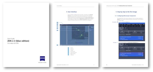
ZEN 2.3 imaging software for microscopy features significant upgrades for imaging with ZEISS research instruments. ZEN 2.3 includes new features and improvements for our next-generation ZEISS Axiocam microscope cameras, ZEISS LSM 800 confocal microscope, and the new ZEISS Axio Observer microscope platform, among others. Make your imaging and user training more efficient with our newest guides, video tutorials and FAQ website!


ZEISS Software Tutorial: Introduction to the ZEN 2.3 3Dxl Module
The software tutorial gives an introduction to the new 3Dxl rendering module available for ZEN (blue) 2.3, powered by arivis.

Can’t see the embedded video? Click here!

ZEISS Software Tutorial: Working with Ultrafast Deconvolution (GPU DCV) in ZEN 2.3
The software tutorial gives an overview of the advanced GPU-based deconvolution module available for ZEN (blue) 2.3.

Can’t see the embedded video? Click here!

ZEISS Software Tutorial: Introduction to Definite Focus.2 in ZEN 2.3
The software tutorial gives an introduction to the Definite Focus.2 functions “Find Surface” and “Lock Focus” in ZEN (blue) 2.3.

Can’t see the embedded video? Click here!

More ZEISS video tutorials are available on our YouTube playlist: Imaging Software for Microscopy


Quick Guide for ZEN 2.3 (blue edition) – First steps with ZEN

ZEN 2.3 Quick Guide

The new Quick Guide for ZEN 2.3 gives users an introduction into the basic functions of ZEN such as the layout of the user interface and the workspace, and provides a step-by-step tutorial for acquisition of the first image. The download is available for free by following this link. Die deutsche Version des Quick Guides ist hier verfügbar


ZEISS FAQ Pages for Light Microscopy

ZEISS Imaging Software for Microscopy

Our new FAQ (Frequently Asked Questions) pages collect various materials and helpful content regarding software and hardware for light microscopy applications, including ZEN and Axiovision guides. Visit the overview here!

For more details about software, services & trainings please contact your ZEISS account manager or use our web contact form: www.zeiss.com/microscopy/contact

You can download your personal copy of the free ZEN lite image viewer at www.zeiss.com/zen-lite

Want to stay updated on our latest products, technology and applications? Subscribe to our newsletter!

Please note that your professional ZEISS confocal, widefield, superresolution or light sheet microscopy workstation is supported by us on the basis of the pre-installed system image, e.g. Windows 7 64bit with ZEN 2.0. Before performing any upgrades to the operating system or ZEN imaging software, please consult with your ZEISS account manager first.

The post ZEN Imaging Software – New Quick Guides & Tutorials Available appeared first on Microscopy News Blog.

Viewing all 43 articles
Browse latest View live